
|
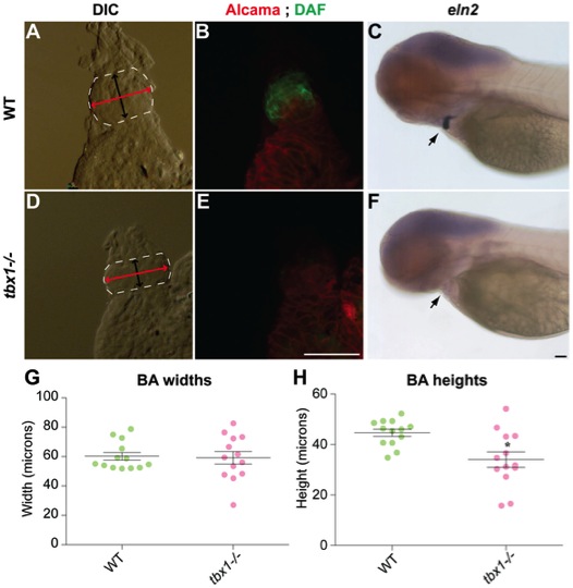
Fig. 4 Bulbous arteriosus defects in tbx1-/- embryos.
Dissected hearts from 72 hpf embryos showing the BA region (A, D) outlined in white, with the bidirectional arrowheads in black and red showing the length and width of BA, respectively. (B, E) The corresponding images showing staining for Alcama antibody (red) and DAF-2DA (green). (C, F) tbx1-/- mutants at the same stage have absent eln2 expression. Black arrows indicate the BA. (G) Quantification of BA width in WT and tbx1-/- mutants, while (H) demonstrates that BA length is reduced in tbx1-/- mutants; * p-value = 0.0045. N = 13 for all measurements. Scale bars: 25 μm.