
|
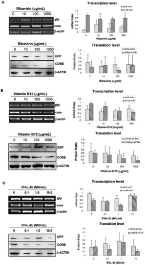
Fig. 6 Verification for the efficiency of the HCV IRES-mediating expression zebrafish model with anti-HCV drugs.
RT-PCR (upper panel) and Western blotting (lower panel) were used for detecting expression of core and gfp in pFL-GIC-injected larvae that were exposed to ribavirin (A) and vitamin B12 (B) drugs at gradient concentrations from 5-dpf to 10 dpf. C. Result of IFNα-2b co-injected with plasmid pFL-GIC. All the larvae were collected at 10-dpf for RT-PCR and Western blotting assays. Untreated larvae were as a control. Both cDNA and protein bands were scanned against β-actin cDNA or β-ACTIN protein respectively for semiquantitative evaluation of core and gfp expression (right histograms).