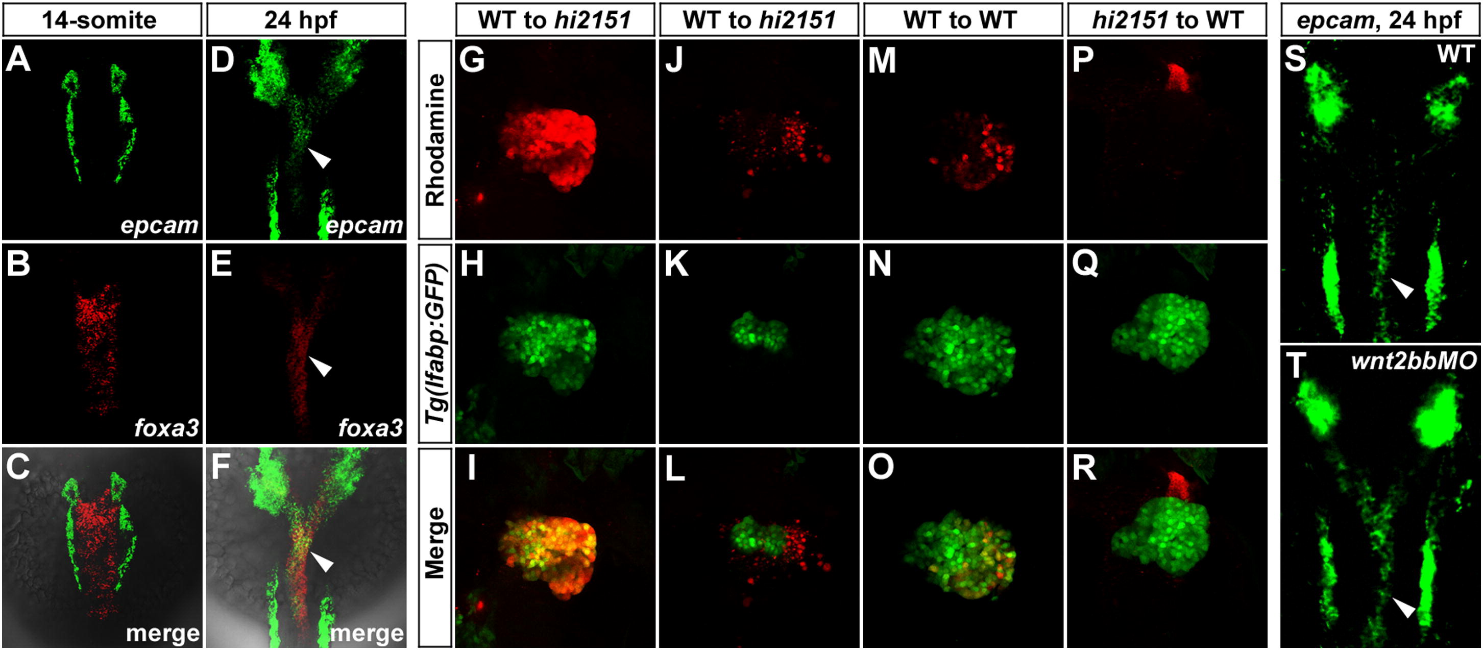
Fig. 3 EpCAM Is Enriched in the Endoderm and Cell Autonomously Modulates Wnt2bb-Induced Hepatic Development(A?F) Double fluorescent in situ hybridizations (FISH) of epcam and foxa3 at the 14-somite stage (A?C) and 24 hpf (D?F). In the endoderm, note that epcam is not detected at the 14-somite stage, but enriched at 24 hpf (arrowheads). foxa3 is an endodermal marker.(G?R) Mosaic analyses using embryos injected with rhodamine-dextran as donors. Only wild-type donor cells massively contributing to the hepatic endoderm (G?I, n = 5), but not those contributing to the surrounding LPM (J?L, n = 8), are able to rescue the reduced liver size in hi2151 mutant acceptors. Transplanted wild-type donor cells (M?O, n = 8), but not hi2151 mutant donor cells (P?R, n = 10), are able to obviously contribute to the liver of wild-type acceptors. Tg(lfabp:GFP) transgenic background is applied in all the donors and acceptors. Transplanted embryos are imaged at 60 hpf.(S and T) As shown by FISH, expression of epcam in the endoderm at 24 hpf (S, 51/51, arrowhead) remains in wnt2bb morphants (T, 48/48, arrowhead).See also Figure S4 and Table S1.
Reprinted from Developmental Cell, 24(5), Lu, H., Ma, J., Yang, Y., Shi, W., and Luo, L., EpCAM Is an Endoderm-Specific Wnt Derepressor that Licenses Hepatic Development, 543-553, Copyright (2013) with permission from Elsevier. Full text @ Dev. Cell

