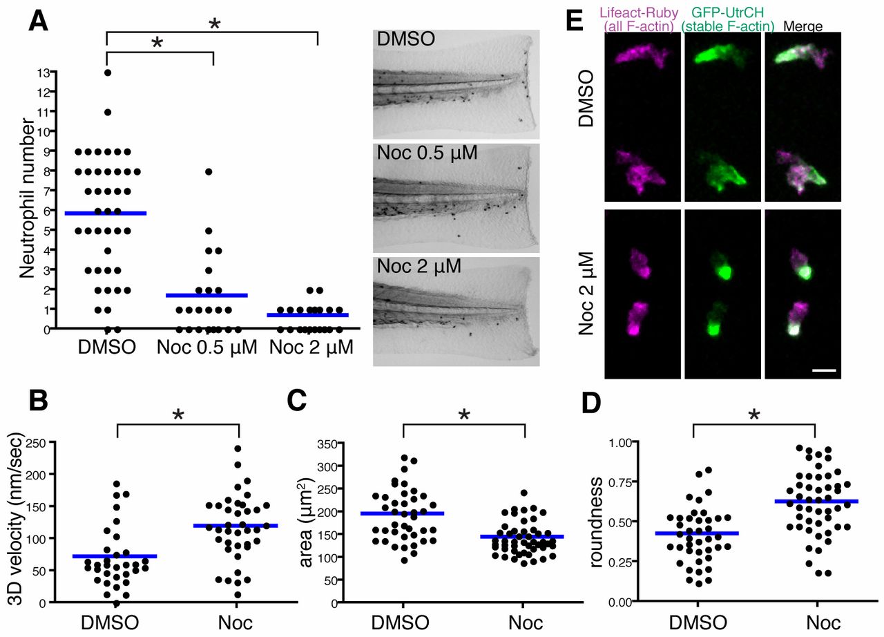
Fig. 3 Microtubule depolymerization impairs neutrophil wound attraction but enhances neutrophil motility and F-actin polarity. (A) Neutrophil recruitment to wounded fins at 30minutes post wounding. Neutrophils were stained with Sudan Black. n = 42 (DMSO), 22 (Noc 0.5μM) and 22 (Noc 2&mu′M). (B?D) Microtubule depolymerization with nocodazole treatment enhances neutrophil 3D motility [(B), n = 32 (DMSO), n = 38 (Noc)], makes cells compact [(C), n = 40 (DMSO), n = 48 (Noc)] and induces a more round morphology [(D), n = 40 (DMSO), n = 48 (Noc)]. Tg(mpx:GFP-UtrCH) was used for live analysis. (E) Nocodazole treatment enhances polarity of F-actin dynamics in neutrophils. Rear localization of stable F-actin detected by GFP-UtrCH is particularly emphasized after microtubule depolymerization. Tg(mpx:GFP-UtrCH) was crossed with Tg(mpx:Lifeact-Ruby). *P<0.05, one-way ANOVA with Dunnett post-test (A) and two-tailed unpaired t-test (B?D). Data in A are representative of at least three separate experiments and three time-lapse movies were analyzed for data in B?D. Scale bar: 10 μm.
Image
Figure Caption
Acknowledgments
This image is the copyrighted work of the attributed author or publisher, and
ZFIN has permission only to display this image to its users.
Additional permissions should be obtained from the applicable author or publisher of the image.
Full text @ J. Cell Sci.

