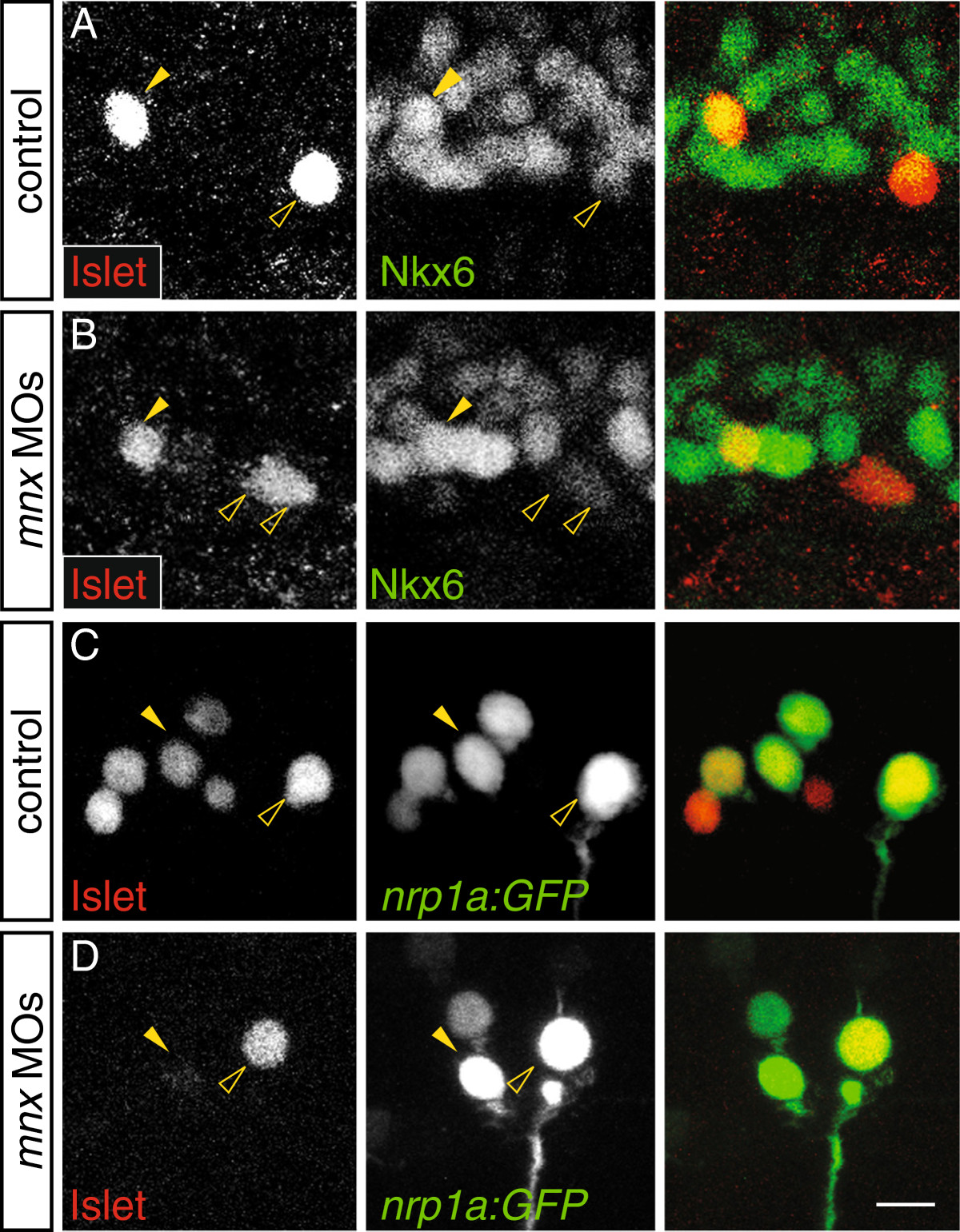
Fig. 6 Mnx proteins maintain the late phase of Islet1 expression in MiPs independently of Nkx6. (A-D) Single spinal hemisegments of control and mnx MO-injected embryos. MiPs (closed arrowheads) and CaPs (open arrowheads) are indicated in each of the single channel panels; right column shows merged channels. (A) At 17 hpf, MiPs in control embryos strongly express Islet and Nkx6 (17/30 Nkx6+ MiPs in seven embryos). CaPs strongly express Islet but weakly express Nkx6. (B) MiPs in mnx MO-injected embryos continue to strongly express Islet and Nkx6 (13/22 Nkx6+ MiPs in five embryos). (C) At 21 hpf in Tg(nrp1a:GFP) control embryos, both MiPs and CaPs express Islet (32/32 Islet+ CaPs, and 31/32 Islet+ in five embryos. (D) In mnx MO-injected Tg(nrp1a:GFP) embryos, CaPs (60/60 Islet+ CaPs in 10 embryos) but not MiPs (19/60 Islet+ MiPs in 10 embryos), strongly express Islet. Scale bar: 20 μm.
Image
Figure Caption
Figure Data
Acknowledgments
This image is the copyrighted work of the attributed author or publisher, and
ZFIN has permission only to display this image to its users.
Additional permissions should be obtained from the applicable author or publisher of the image.
Full text @ Neural Dev.

