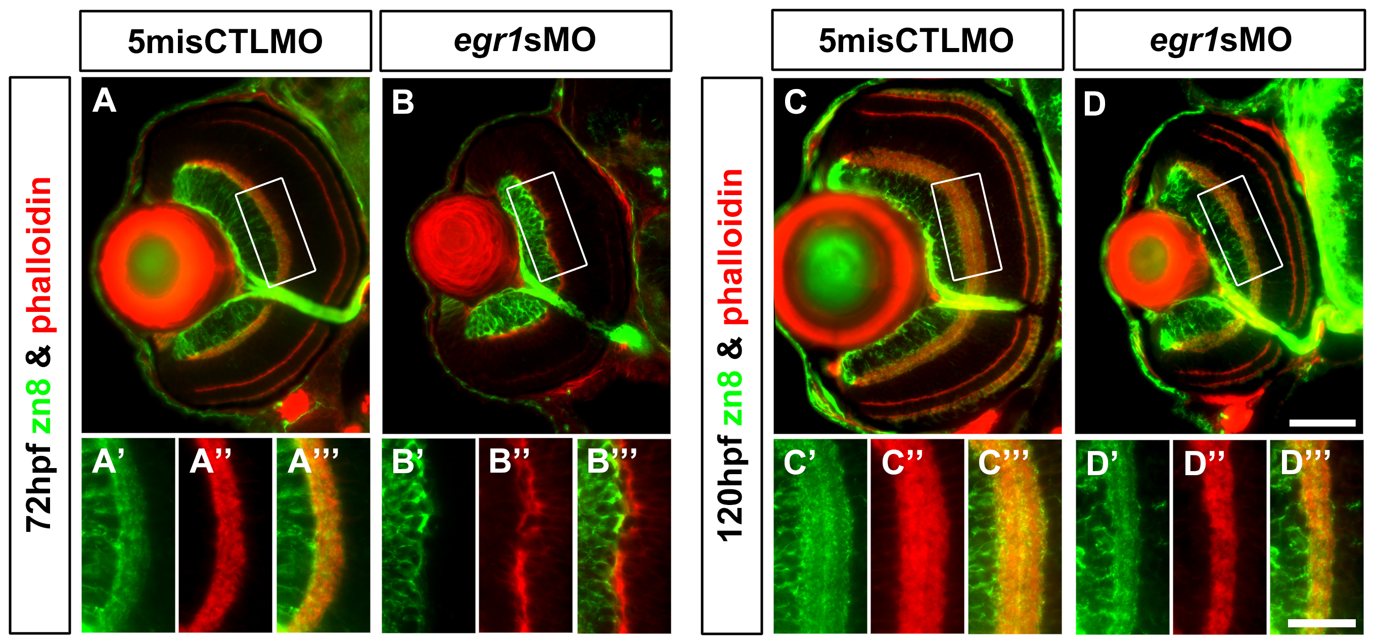
Fig. 5 Immunohistochemical analysis of the GCs in the Egr1-morphant retinas.
Immunohistochemical analysis of the GCs in the controls (5misCTLMO) and Egr1 morphants (egr1sMO) was performed by anti-zn8 (zn8; green) at 72 hpf (A & B) and 120 hpf (C & D). Phalloidin (red) was used as a counterstain to highlight the plexiform layers. A whole-eye section is shown at the top for each condition, while the magnified view of a selected region (white box) on the dorsal side of the optic nerve is shown at the bottom. The analysis has indicated that Egr1 knockdown suppressed the early dendritic outgrowth of GCs into the IPL at 72hpf (B), which was irregular at this stage. In addition, the cell number per retinal area was not different between the two groups. This defect was largely resolved by 120 hpf, despite the IPL was still thinner as shown in Figure 3. This suggests that there were still defects in differentiation of cells that projected neurites into the IPL. One possible cause of the defect is the differentiation problem of ACs as shown in Figure 4. See text, Table 1 and 2 for further discussion. For the whole-eye sections, the lens is on the left and dorsal is up. Scale bar = 50 Ám for the whole-eye sections and 25 Ám for the selected regions.

