
|
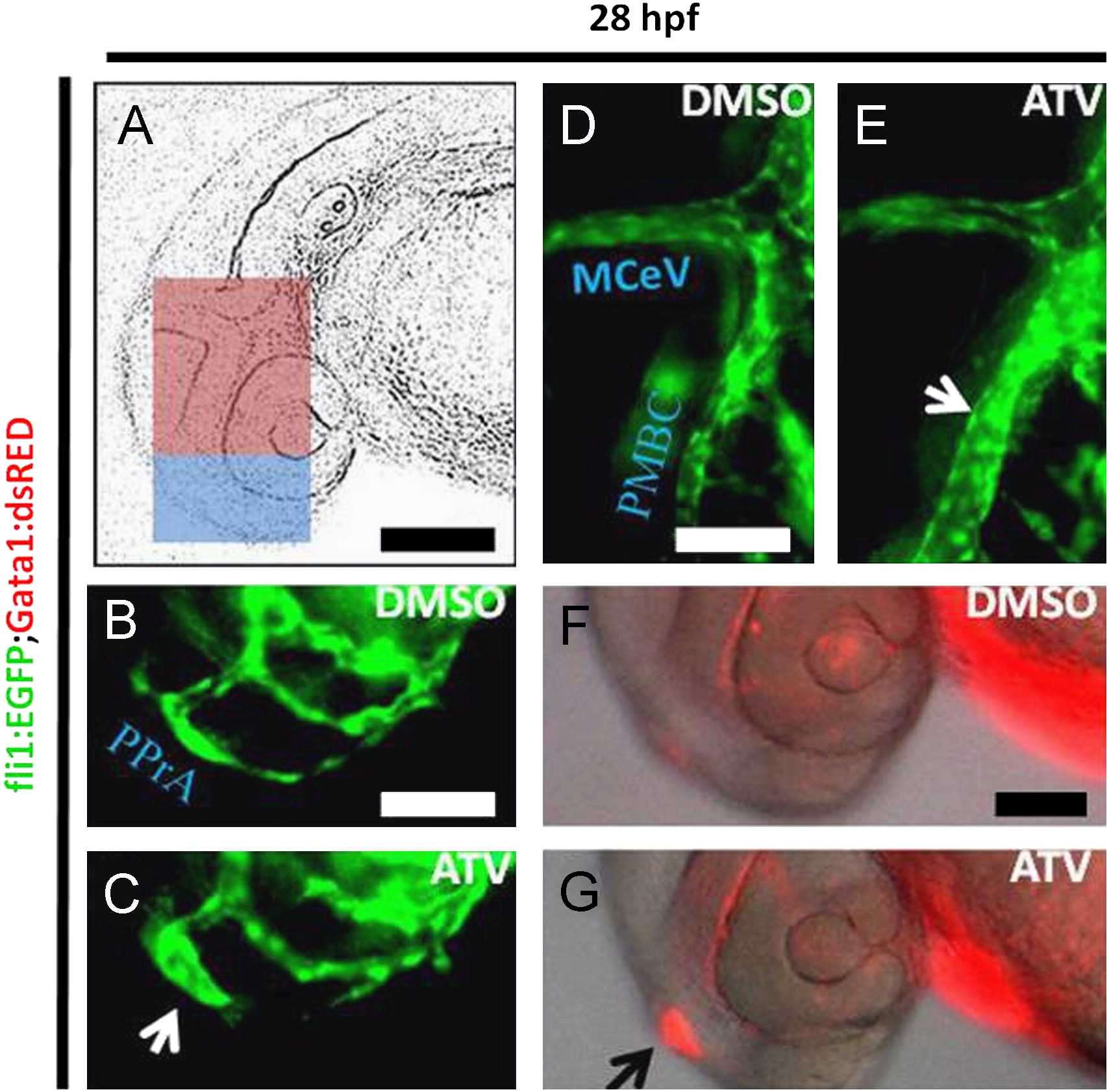
Fig. 1 Pharmacological inhibition of the HMGCR pathway induces cerebral-vascular defects in developing zebrafish. (A) Representative schematic diagram of a 28 hpf zebrafish embryo, with the blue boxed area denoting the forebrain and the pink boxed area showing the midbrain regions. Scale bar=200 μm. ((B)–(G)) Representative photomicrographs of Tg(fli1:EGFP);(gata-1:DsRed) embryos soaked in DMSO or 0.5 mg/L ATV and imaged at 28 hpf. Abbreviations: PMBC, primordial midbrain channel; MCeV, middle cerebral vein; PPrA, primitive prosencephalic artery. White arrows indicate the abnormally dilated vessels in the forebrain and midbrain, whereas the black arrow denotes area of stagnant erythrocyte accumulation in the head. Scale bar=40 μm.
Reprinted from Developmental Biology, 373(2), Eisa-Beygi, S., Hatch, G., Noble, S., Ekker, M., and Moon, T.W., The 3-hydroxy-3-methylglutaryl-CoA reductase (HMGCR) pathway regulates developmental cerebral-vascular stability via prenylation-dependent signalling pathway, 258-266, Copyright (2013) with permission from Elsevier. Full text @ Dev. Biol.