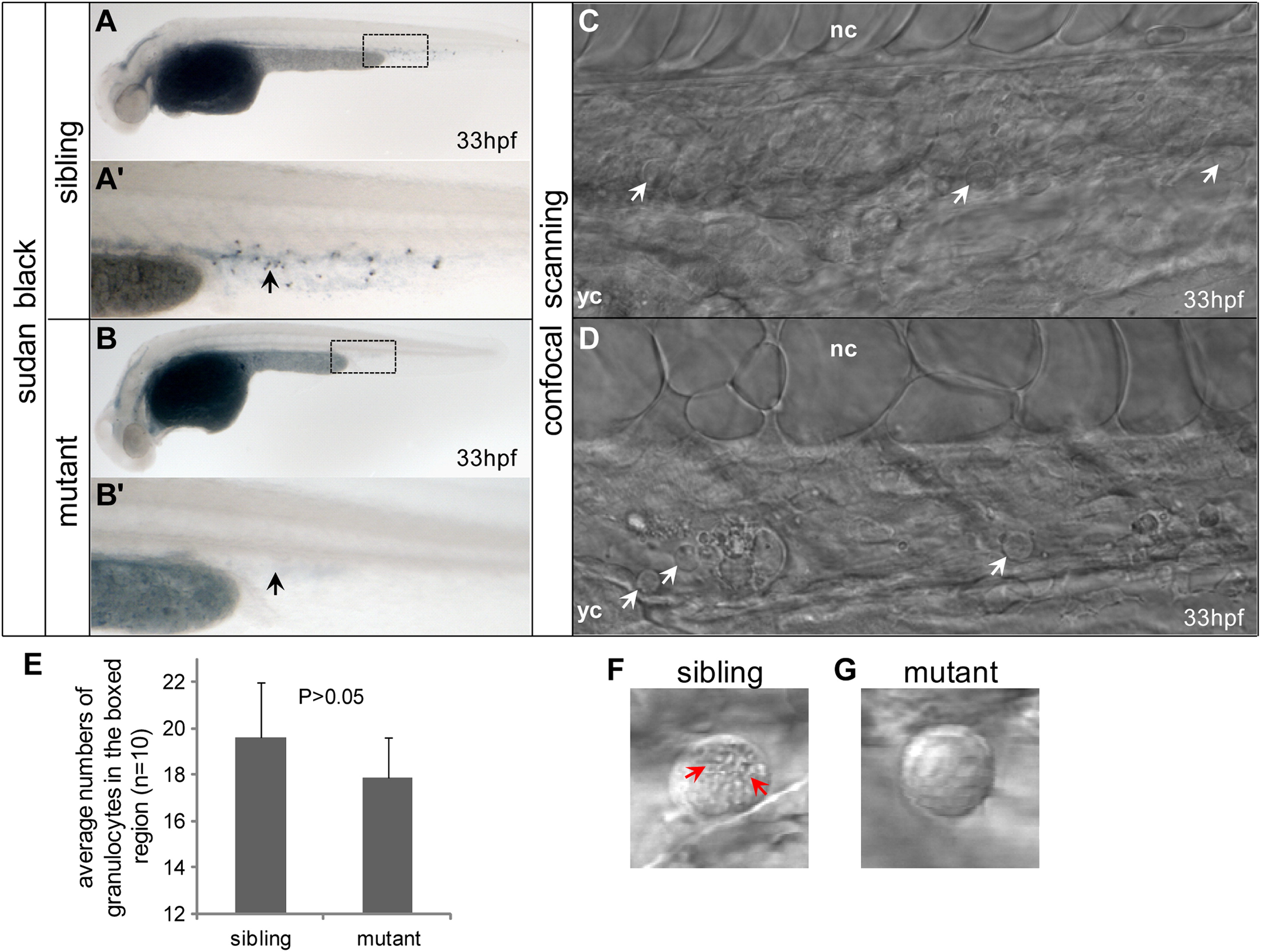
Fig. 6 Disruption of plcg1 plays an important role in terminal maturation of granulocytes. ((A) and (B), (A′) and (B′)) Sudan black staining of plcg1 mutants. In the mutants, the staining is significantly lower ((B) and (B2), black arrow) than that in wild-type siblings ((A) and (A′), black arrow). ((C) and (D)) Confocal scanning of live wild-type sibling and mutant embryos. The equivalent scanned area is shown ((A) and (B), dotted squares). In one scanning layer, some round and big cells similar to granulocytes could be detected and the numbers of these group cells are equal between wild-type siblings ((C), white arrows) and mutants ((D), white arrows). notochord: nc. yolk sac: yc. (E) Average numbers of granulocytes in the boxed region between siblings and mutant embryos. ((F) and (G)) Higher magnification of granulocytes between siblings and mutant embryos. Red arrows represent granules of granulocyte (F). (For interpretation of the references to color in this figure legend, the reader is referred to the web version of this article.)
Reprinted from Developmental Biology, 374(1), Jing, C.B., Chen, Y., Dong, M., Peng, X.L., Jia, X.E., Gao, L., Ma, K., Deng, M., Liu, T.X., Zon, L.I., Zhu, J., Zhou, Y., and Zhou, Y., Phospholipase c gamma-1 is required for granulocyte maturation in zebrafish, 24-31, Copyright (2013) with permission from Elsevier. Full text @ Dev. Biol.

