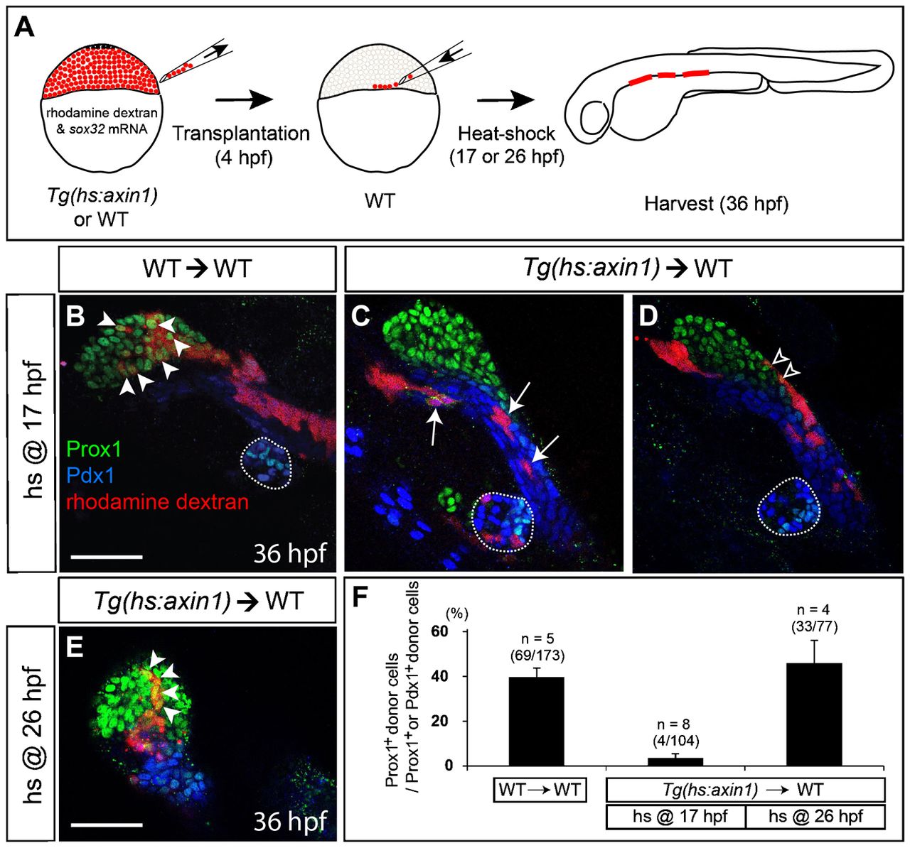
Fig. 3 Endodermal cells with Wnt/β-catenin signaling repressed fail to contribute to the liver.
(A) Cartoons illustrating transplantation experiments. Wild-type or Tg(hs:axin1) embryos were co-injected with sox32 mRNA and rhodamine dextran at the 1- to 4-cell stage. Donor cells from the injected embryos were transplanted into a wild-type embryo at 4hpf. The transplants were heat-shocked at 17 (B?D) or 26 (E) hpf, harvested at 36hpf, and processed for immunostaining. (B,C) Wild-type donor cells expressed Prox1 (green) in the liver-forming region (B, arrowheads), whereas Axin1-overexpressing donor cells heat-shocked at 17hpf expressed Pdx1 (blue) but not Prox1 (C, arrows). (D) A few Axin1-overexpressing donor cells were located at the margin of the liver-forming region (open arrowheads). (E) Axin1-overexpressing donor cells heat-shocked at 26hpf expressed Prox1 and were located in the liver-forming region (arrowheads). (F) Quantification of the transplantation experiments. Percentages of Prox1+ donor cells relative to Prox1+ or Pdx1+ donor cells are shown. Error bars represent the standard deviation; n indicates the number of embryos. Dotted lines outline the dorsal pancreas. Ventral views, anterior up (B?E). Scale bars, 50Ám.

