
|
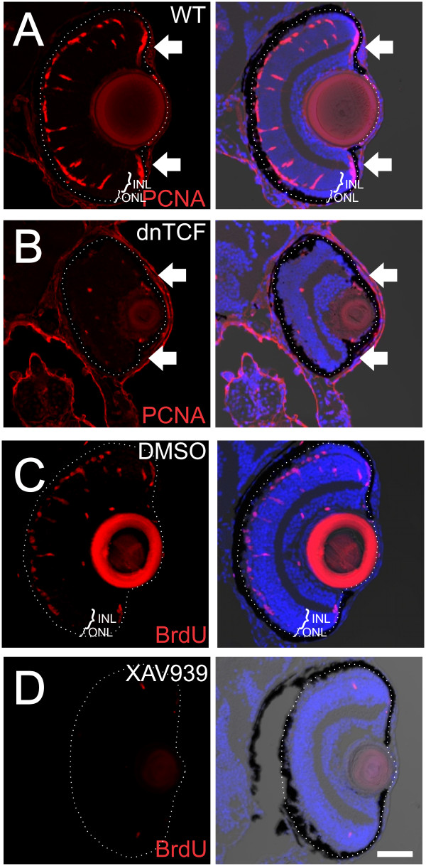
Fig. 8 Inhibition of Wnt signaling with dnTCF or XAV939 inhibits injury-induced proliferation. (A, B) dnTCF zebrafish larvae and wild-type siblings were given an intense light lesion at 6 dpf and heat shocked at 39.5�C for 1 h at 1 dpl and again at 2 dpl then fixed at 3 dpl. In wild-type siblings, PCNA-labeled proliferative cells are in the CMZ (arrows), INL, and ONL at 3 dpl (A), while dnTCF zebrafish have few proliferative cells in either the retina or the CMZ (B). (C, D) Wild-type zebrafish lesioned as in (A, B) were treated with 0.15% DMSO or 15 �M XAV939, with 2.5 mM BrdU present continuously in the media until fixation at 3 dpl. In DMSO-treated fish, BrdU-positive cells are in the INL and ONL (C), while XAV939-treated fish have few proliferative cells in either the INL or ONL (D). Scale bar = 50 �m.