Image
Figure Caption
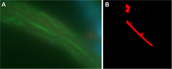
Fig. 2 Immunohistochemical staining of skeletal muscle biopsy from the back side of zebrafish with MAb Tit1 5 H1.1. (A) Cryoslice of zebrafish skeletal muscle immunostained with MAb Tit1 5 H1.1, fixed with 4% PFA, specific staining (green) with Alexa 488, obj.100x. (B) Mechanically separated skeletal muscle fibre of zebrafish, fixed with 4% PFA, immunostained with MAb Tit1 5 H1.1, specific staining (red) with Alexa 594, obj.100x. Slices were embedded with Prolong Gold anti-fade reagent.
Figure Data
Acknowledgments
This image is the copyrighted work of the attributed author or publisher, and
ZFIN has permission only to display this image to its users.
Additional permissions should be obtained from the applicable author or publisher of the image.
Full text @ Cell Div.

