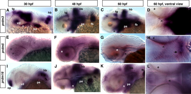
Fig. 1 Expression of different prdms in developing zebrafish embryos. In situ hybridization (ISH) using prdm3, 5, and 16 at 30-hpf (A, E, I), 48-hpf (B, F, J), and 60-hpf stages showing lateral (C, G, K) and (D, H, L) ventral views. Note the partially overlapping expression of prdm3 and prdm16. A: Lateral view of whole mount embryo showing the prdm3 expression in the tegmentum, ventral diencephalon, neurocranium, hindbrain, pharyngeal arches at 30 hpf. At 48 hpf (B) and 60 hpf (C, D), prdm3 gradually increases and is highly expressed in pharyngeal arches, and is also expressed in pectoral fin buds, neurocranium, hindbrain, and tegmentum. E?H: prdm5 is not specifically expressed at 30 hpf, but is expressed at a low level in the area behind the eye and neurocranium at 48 and 60 hpf. The arrow in H points to the forming stomodeum in which prdm5 is expressed. I,J: From 30 to 48 hpf, prdm16 expression gradually increases in pharyngeal arches, as well as in neurocranium, pectoral fin buds, hindbrain, and olfactory placode. K,L: At 60 hpf, prdm16 expression decreases in the pharyngeal arches and hindbrain, but is still expressed at a significant level. Anterior is to the left in all panels. e, eye; fb, fin buds; hb, hindbrain; n, neurocranium; op, olfactory placode; pa, pharyngeal arch; tg, tegmentum; vd, ventral diencephalon.
Image
Figure Caption
Figure Data
Acknowledgments
This image is the copyrighted work of the attributed author or publisher, and
ZFIN has permission only to display this image to its users.
Additional permissions should be obtained from the applicable author or publisher of the image.
Full text @ Dev. Dyn.

