Image
Figure Caption
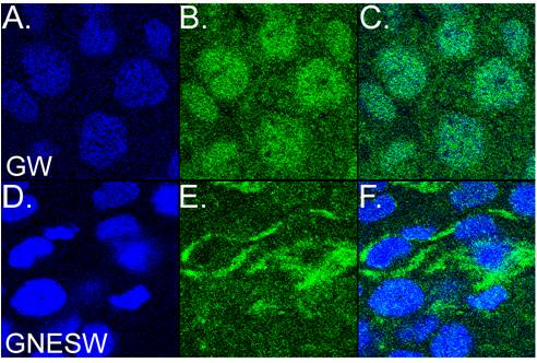
Fig. S1 The GFPNESWdr68 fusion redistributes to the cytoplasm in zebrafish. A, D) DAPI stained cell nuclei. B, E) GFP fusion proteins. C, F) Overlays of blue and green channels. A?C) Moderate nuclear enrichment of GFPWdr68 (GW) in cells of late epiboly stage zebrafish embryos. D?F) Predominant nuclear exclusion of GFPNESWdr68 (GNESW) in cells of late epiboly stage zebrafish embryos.
Acknowledgments
This image is the copyrighted work of the attributed author or publisher, and
ZFIN has permission only to display this image to its users.
Additional permissions should be obtained from the applicable author or publisher of the image.
Full text @ PLoS One

