
|
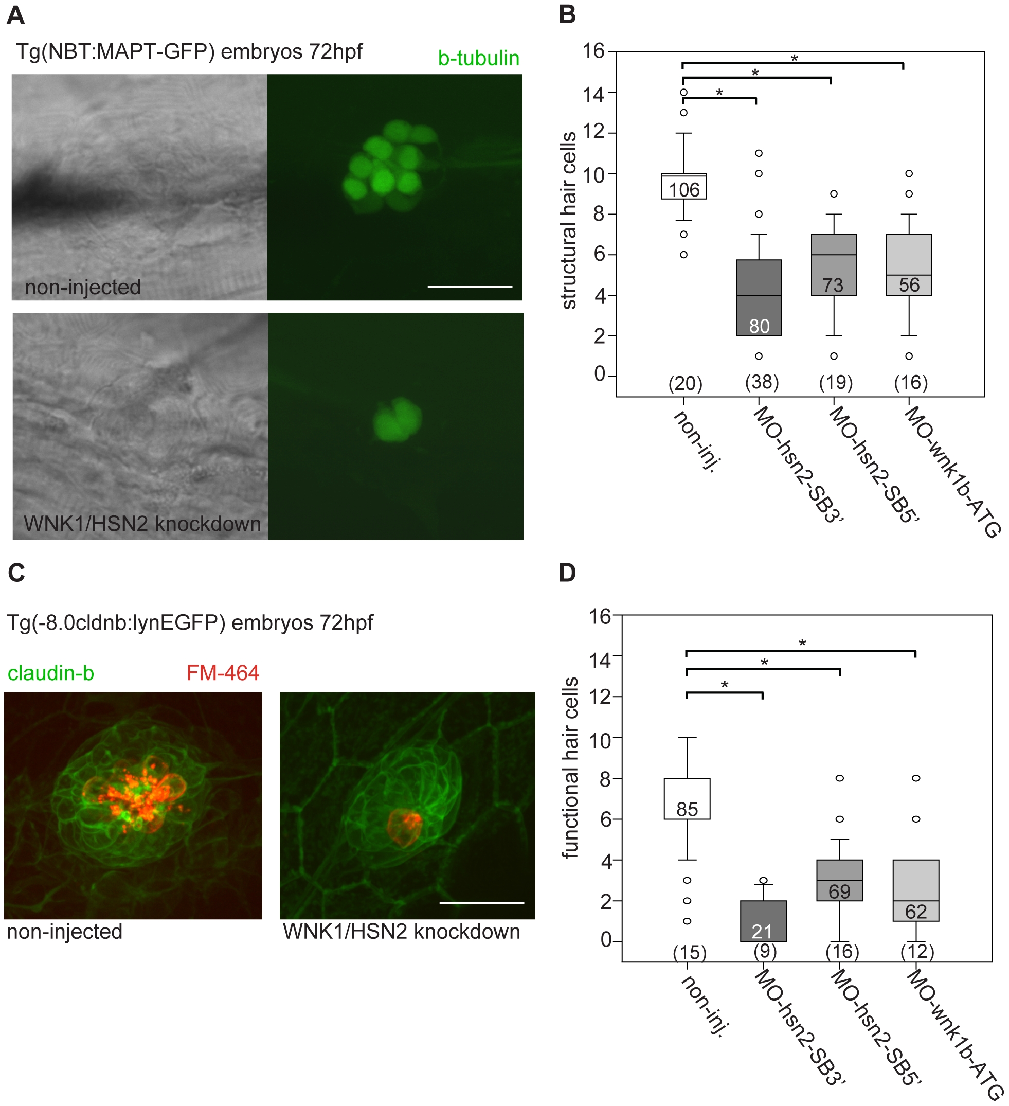
Fig. 3
WNK1/HSN2 knockdown leads to abnormal neuromast development.
A) The number of structural hair cells was assessed using transgenic embryos expressing GFP under the beta-tubulin promoter, revealing only the neuronal hair cells within PLL neuromasts. C) The number of functional hair cells was assessed by using transgenic embryos expressing GFP under the claudin-b promoter, rendering the neuromast fluorescent. The functional hair cells were revealed by incubation in the styryl dye FM-464FX, shown in red. B, D) Hair cells were counted for each PLL neuromast and totals were tabulated in box plots showing that WNK1/HSN2 knockdown embryos have a significantly lower number of structural and functional hair cells within their neuromasts when compared with non-injected embryos. The knockdown embryos presented in (A) and (C) are representative results at 72hpf obtained from MO-hsn2-SB32 injection. The number of neuromasts counted per condition is indicated in the boxes and the total number of embryos obtained per condition is indicated in parenthesis at the bottom of the box plots. Scale bar: 20 μm.