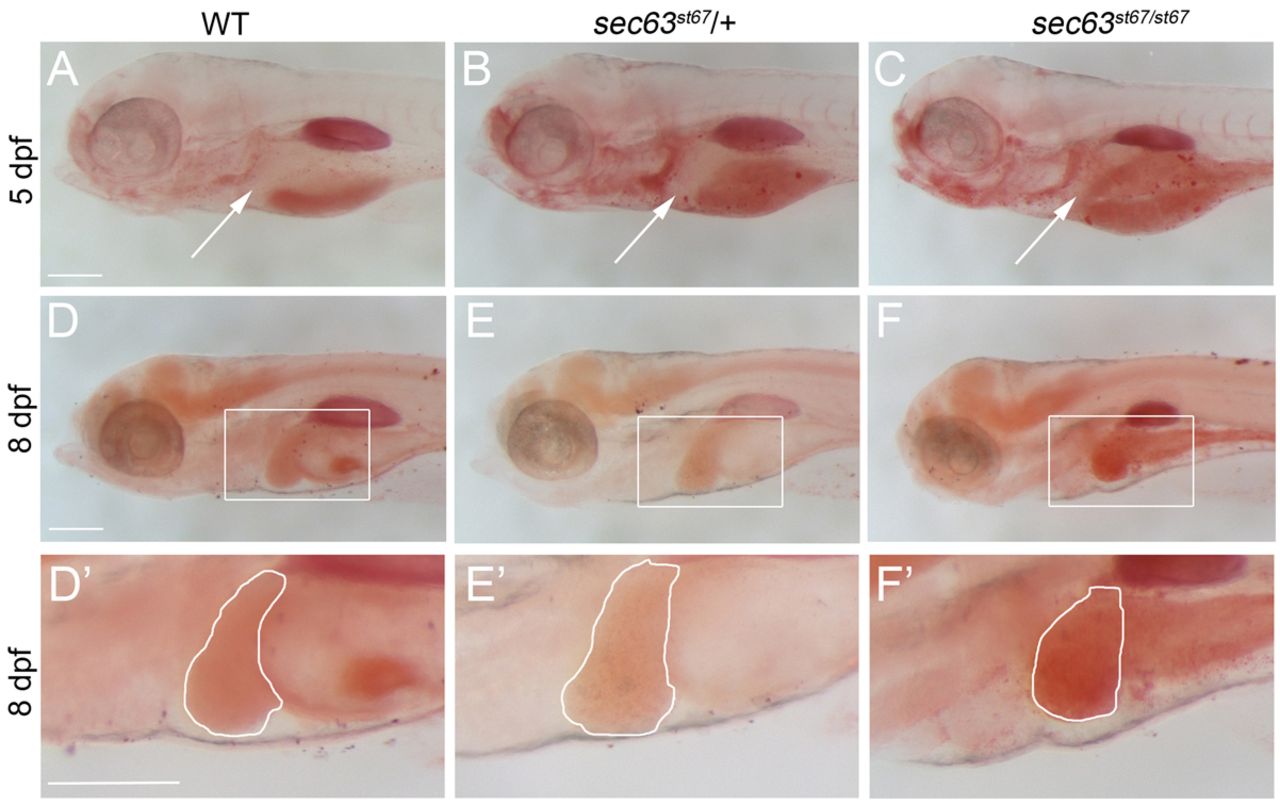
Fig. 7 sec63st67 mutants develop liver steatosis. (A–F2) Lateral views of larvae stained with Oil Red O of the indicated genotypes and at the indicated developmental stages. (A–C) Arrows indicate the location of the liver in 5 dpf larvae. sec63st67 mutant livers (C, n=7) are indistinguishable from wild-type (A, n=6) or heterozygous (B, n=12) livers. (D–F) Boxed regions denote the areas enlarged in D′–F′. (D′–F′) Outlines denote the liver in 8 dpf larvae. sec63st67 mutant livers (F,F′, n=8) show stronger Oil Red O stain than wild-type (D,D′, n=14) or heterozygous (E,E′, n=34) livers. Scale bars: 200 μm.
Image
Figure Caption
Figure Data
Acknowledgments
This image is the copyrighted work of the attributed author or publisher, and
ZFIN has permission only to display this image to its users.
Additional permissions should be obtained from the applicable author or publisher of the image.
Full text @ Dis. Model. Mech.

