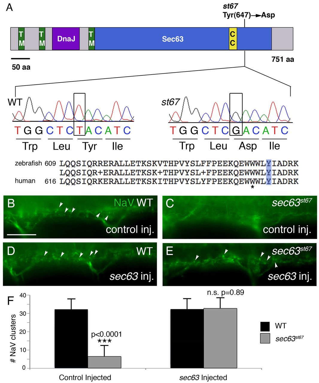
Fig. 4 st67 disrupts zebrafish sec63. (A) Representation of Sec63 showing functional domains and the location of the lesion in the st67 mutation. TM, transmembrane domain; DnaJ, DnaJ domain; Sec63, Sec63 domain; CC, coiled-coil region. Also shown are sequence traces from homozygous wild-type and st67 mutant larvae. The st67 mutation changes a conserved tyrosine to an aspartic acid in the Sec63 domain. A comparison of zebrafish and human Sec63 amino acid sequence in the vicinity of the st67 mutation is also shown. The light blue box indicates the location of the lesion in st67 zebrafish mutants. The asterisk marks the position of a SEC63 mutation identified in human patients with PCLD (W651G) (Waanders et al., 2010). (B–E) Representative images of antibody-stained preparations of the spinal cord of larvae of the indicated genotypes and injection treatments at 72 hpf. (B) Siblings injected with control solution show normal NaV clustering (arrowheads). (C) st67 mutants injected with control solution show aberrant NaV clustering. (D,E) Siblings and st67 mutants injected with 150 pg of synthetic sec63 mRNA show normal NaV clustering. (F) Quantification of the total number of NaV puncta in two hemisegments (<200 μm) of ventral spinal cord of sibling (black bars) and st67 mutant larvae (gray bars) at 72 hpf following the indicated injection regimes. The P values for unpaired t-test comparisons (two-tailed) are shown; error bars indicate s.d. Sample sizes: 9 control-injected siblings. 6 control-injected mutants, 12 sec63-injected siblings and 11 sec63-injected mutants. Scale bar: 20 μm (B–E).
Image
Figure Caption
Figure Data
Acknowledgments
This image is the copyrighted work of the attributed author or publisher, and
ZFIN has permission only to display this image to its users.
Additional permissions should be obtained from the applicable author or publisher of the image.
Full text @ Dis. Model. Mech.

