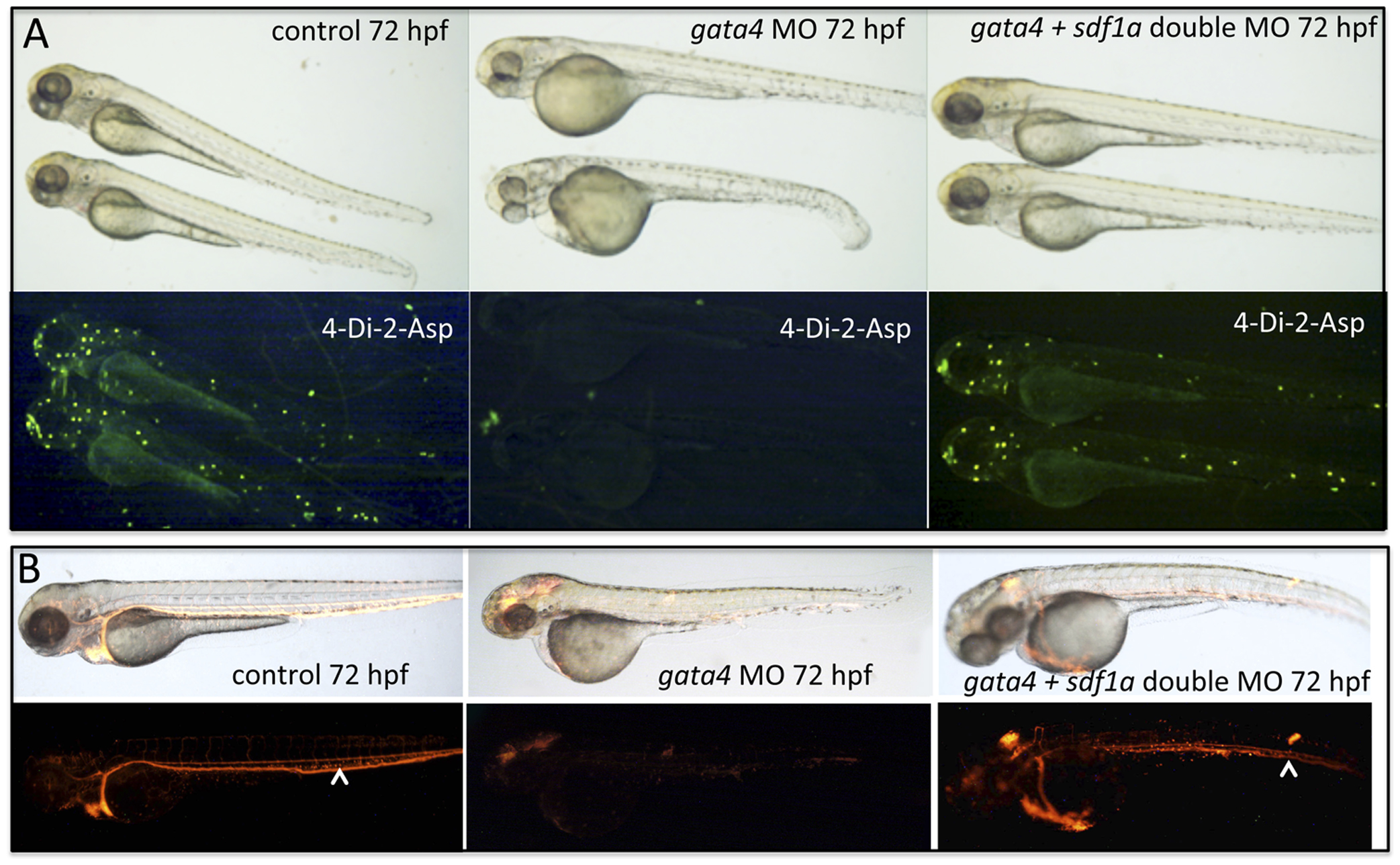
Fig. 7 Depletion of sdf1a rescues the neuromast and circulation defects in the gata4 morphant.
A: Shown are representative embryos at 72 hpf (n >50, multiple independent experiments) following incubation with 4-Di-2-Asp to detect neuromasts. For comparison with control embryos (left panels) embryos were injected with the gata4 morpholino and half the clutch was left alone (middle panels), while the other half was subsequently injected with the sdf1a morpholino (right panels). The lower panels are the fluorescence images of the corresponding brightfield panels. The examples shown are representative of the rescue for the lateral line in 34% of co-injected embryos (using 0.25 ng of the sdf1a morpholino). B: Shown are representative embryos at 72 hpf (n >50, multiple independent experiments) derived from the tg(gata1:dsRed) reporter line. For comparison with control embryos (left panels) embryos were injected with the gata4 morpholino and half the clutch was left alone (middle panels), while the other half was subsequently injected with the sdf1a morpholino (right panels). The lower panels are the fluorescence images of the corresponding brightfield panels. The rescue of circulation into the CHT plexus in the tail was observed in 82% of co-injected embryos (using 2 ng of the sdf1a morpholino). White arrowheads indicate the caudal vein in the CHT. Views are lateral, anterior to the left.

