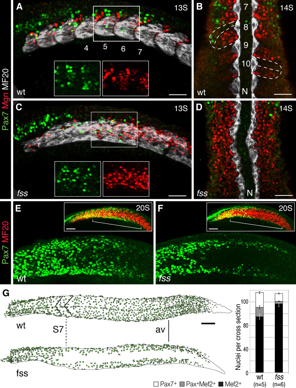
Fig. 2 fss/tbx6 is required for the central dermomyotome development in the posterior trunk and tail.
(A–D) Pax7 (green), Myogenin (Mgn, red), and MF20 (MyHC, white) expression in the anterior trunk (A,C; confocal z-stack, insets show higher magnification) and the posterior trunk (B,D) of wild type sibling (A,B) and fss/tbx6 mutant (C,D) at 13–14S stage. N, Notochordd, numbers indicate somite number. Wild type siblings express all three markers within distinct compartments of the somites (dashed lines in B). In fss/tbx6 mutants, Pax7 expressing central dermomyotome cells are restricted to the anterior trunk; the central posterior trunk shows more cells expressing Mgn. (E,F) Pax7 (green) and MF20 (MyHC, red) expression in wild type sibling (E) and fss/tbx6 mutant (F) embryo at 20S stage. Boxes show flattened confocal z-stacks of the entire trunk. In the posterior trunk Pax7 is initially expressed in cells of the dorsal and ventral dermomyotome; central Pax7 expression is delayed in wild type sibling (E), but absent in fss/tbx6 mutant (F). (G) Tracing of Pax7+ dermomyotome nuclei in representative wild type sibling and fss/tbx6 embryos at the 24h stage. In this mutant embryo, the central dermomyotome deficit/defect in fss/tbx6 mutants starts at trunk levels correlating to somite 7 in the wild type sibling (dashed line, S7). Graph showing number of Pax7+, Mef2+ and differentiating (Pax7+/Mef2+) cells (mean±s.e.m.) on sections at anal vent (av) level in wild type siblings and fss/tbx6 mutants. Scale bars: 50μm (A–D), 100μm (E–G).

