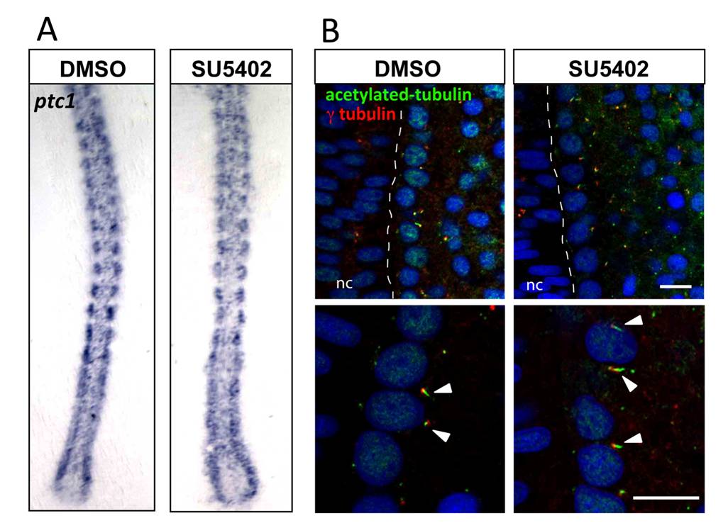
Fig. S2 Hedgehog and primary cilia are not affected by FGF signaling inhibition. (A) ptc1 expression is similar in DMSO and in SU5402 treated embryos at 13-somite, as determined by in situ hybridization. Flat mounted embryos, dorsal view, anterior towards the top. (B) Acetylated-tubulin and γ-tubulin expression in 13-somite embryos after DMSO or SU5402 treatment. Number and length of primary cilia of the adaxial cells in the presomitic mesoderm (arrow heads) are unaffected by SU5402 treatment. Pictures are single confocal scans, dorsal view. Dashed line shows the limit between the notochord (nc) and the presomitic mesoderm. Adaxial cells are adjacent to the notochord. Scale bars: 10 μm.
Image
Figure Caption
Acknowledgments
This image is the copyrighted work of the attributed author or publisher, and
ZFIN has permission only to display this image to its users.
Additional permissions should be obtained from the applicable author or publisher of the image.
Full text @ PLoS Genet.

