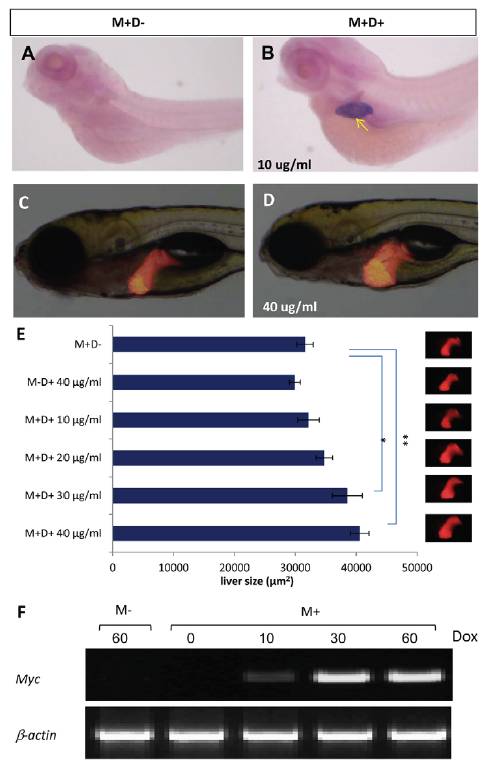
Fig. 1 Inducible Myc expression and liver overgrowth in TO(Myc) fry. (A,B)Liver-specific Myc expression in TO(Myc) fry. Myc transgenic fry were treated with 10 μg/ml Dox from 2 dpf for two days and liver-specific Myc expression was confirmed by whole mount in situ hybridization at 4 dpf, as indicated by a yellow arrow. An untreated control is shown in A. (C,D)In vivo detection of liver overgrowth in TO(Myc) fry. Double transgenic fry from a cross of TO(Myc) and LiPan zebrafish were treated with 40 μg/ml Dox and livers were observed by RFP expression (D). An untreated control is shown in C. (E)Dosedependent growth of liver in TO(Myc) fry. TO(Myc)/LiPan double transgenic fry were treated with increasing concentrations of Dox; liver imaging is shown in the pictures on the right. 2D measurement of liver areas were performed using ImageJ as described previously (Huang et al., 2012) and the quantitative data are shown on the left. The groups significantly different from the control group (M+D-) by Student′s t-test are indicated: *P<0.05; **P<0.01. (F)Dose-dependent induction of Myc mRNAs in adult livers as detected by RT-PCR. Concentrations of Dox are indicated at top of each lane. M+, TO(Myc) fish; M-, non-transgenic siblings; D+, presence of doxycycline; D-, absence of doxycycline. B-actin transcripts served as loading controls for RT-PCR.
Image
Figure Caption
Figure Data
Acknowledgments
This image is the copyrighted work of the attributed author or publisher, and
ZFIN has permission only to display this image to its users.
Additional permissions should be obtained from the applicable author or publisher of the image.
Full text @ Dis. Model. Mech.

