
Image
|
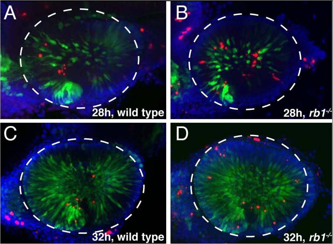
Figure Caption
Fig. S1 rb1te226a retinas show increased apoptosis. Retinas removed from wild type (A, C, E) or rb1te226a; ath5:gfp embryos (B, D, F) at 28 (A?B), 32 (C?D), or 36 hpf (E?F). Retinas labeled anti-GFP (green), TUNEL (red), and counterstained with DAPI (blue). Lateral view of maximum intensity projection of confocal z-stacks. White dashed circle outlines retina. Anterior to the left, dorsal to the top of each panel. (G) Mean number of TUNEL positive nuclei per retina. Error bars denote SEM. *p<0.01; one-way ANOVA. N retinas shown at base of bar graphs. Scale bar = 50 μm.
Acknowledgments
This image is the copyrighted work of the attributed author or publisher, and
ZFIN has permission only to display this image to its users.
Additional permissions should be obtained from the applicable author or publisher of the image.
Full text @ PLoS Genet.