
Image
|
Figure Caption
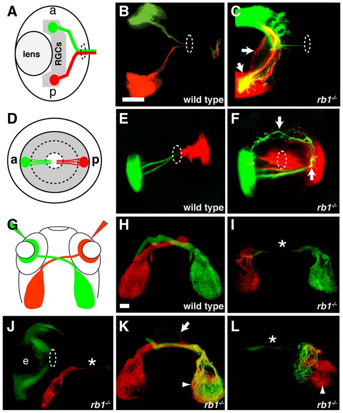
Fig. 7 Intraretinal and midline RGC axon pathfinding errors in rb1te226a mutants.
(A-C) Dorsal, intraretinal views of DiO (green) labeled anterior and DiI (red) labeled posterior RGCs. (D-F) Lateral, intraretinal views of DiO/DiI labeled RGCs. (G-L) Dorsal views of retinotectal projection labeled by whole retina fills with DiI/DiO. Eyes removed in H-I, K-L. Dashed circles indicate retinal exit point. Arrows mark misprojecting axons. Asterisk shows optic nerve hypoplasia. Arrowheads mark ipsilateral tectal innervation. Abbreviations: (a) anterior, (p) posterior, (e) eye. Scale bar = 50 μm.
Figure Data
Acknowledgments
This image is the copyrighted work of the attributed author or publisher, and
ZFIN has permission only to display this image to its users.
Additional permissions should be obtained from the applicable author or publisher of the image.
Full text @ PLoS Genet.