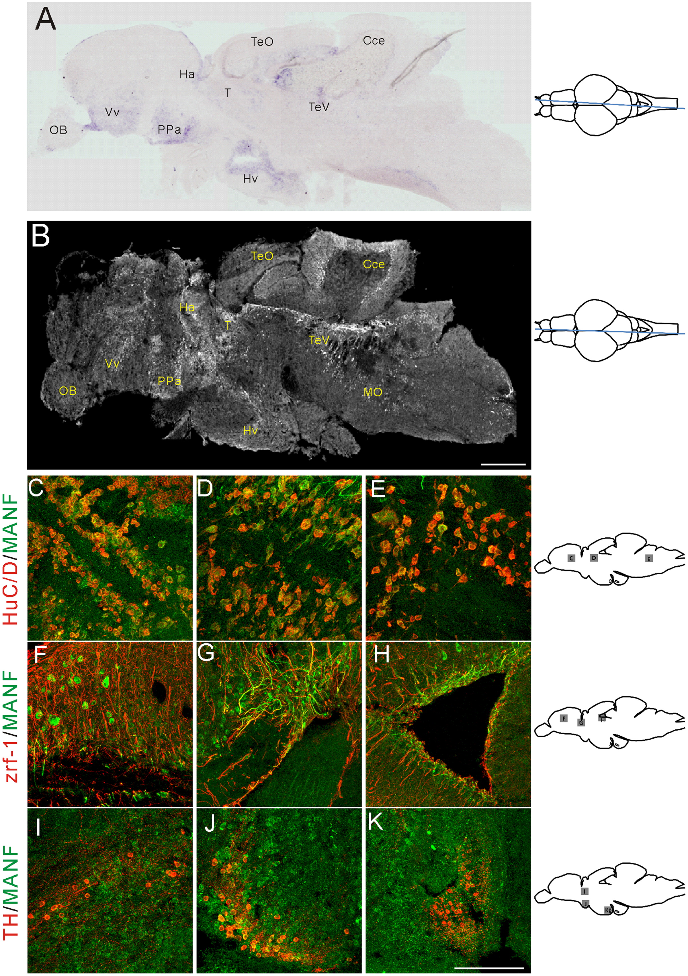
Fig. 2
Fig. 2 In situ hybridization mRNA analysis and immunocytochemistry of MANF expression in adult zebrafish brain. (A) A sagittal view shows manf mRNA in an adult brain section. Expression can be seen in ventral telencephalic, preoptic, ventral thalamic, pretectal, dorsal thalamic, and hypothalamic regions. (B) A sagittal view of the brain shows immuoreactivity for MANF protein. Expression is shown in telencephalic, preoptic, thalamic, optic tectal, cerebellar and rhombencephalic ventricular regions. MANF antibody co-immunostaining with HuC/D antibody in ventral telencephalic (C), dorsal thalamic (D), and rhombencephalic ventricular regions (E). Co-immunostaining with zrf-1 in dorsal telencephalon (F), ventral thalamic region (G), and telencephalic ventricle (H). Co-immunostaining with TH1 in ventral thalamic area (I), preoptic complex (J), and in caudal hypothalamus (K). MANF staining is shown in green; HuC/D, zrf-1 and TH staining are shown in red. Cce: cerebellar corpus. Ha: habenula. Hv: ventral zone of periventricular hypothalamus. OB: olfactory bulb. PPa: parvocellular preoptic nucleus, anterior part. T: thalamus. TeO: tectum opticum. TeV: tectal ventricle. Vv: ventral nucleus of ventral telencephalic area. Scale bar=300 μm in (B), 100 μm in (K).
Reprinted from Developmental Biology, 370(2), Chen, Y.C., Sundvik, M., Rozov, S., Priyadarshini, M., and Panula, P., MANF regulates dopaminergic neuron development in larval zebrafish, 237-249, Copyright (2012) with permission from Elsevier. Full text @ Dev. Biol.

