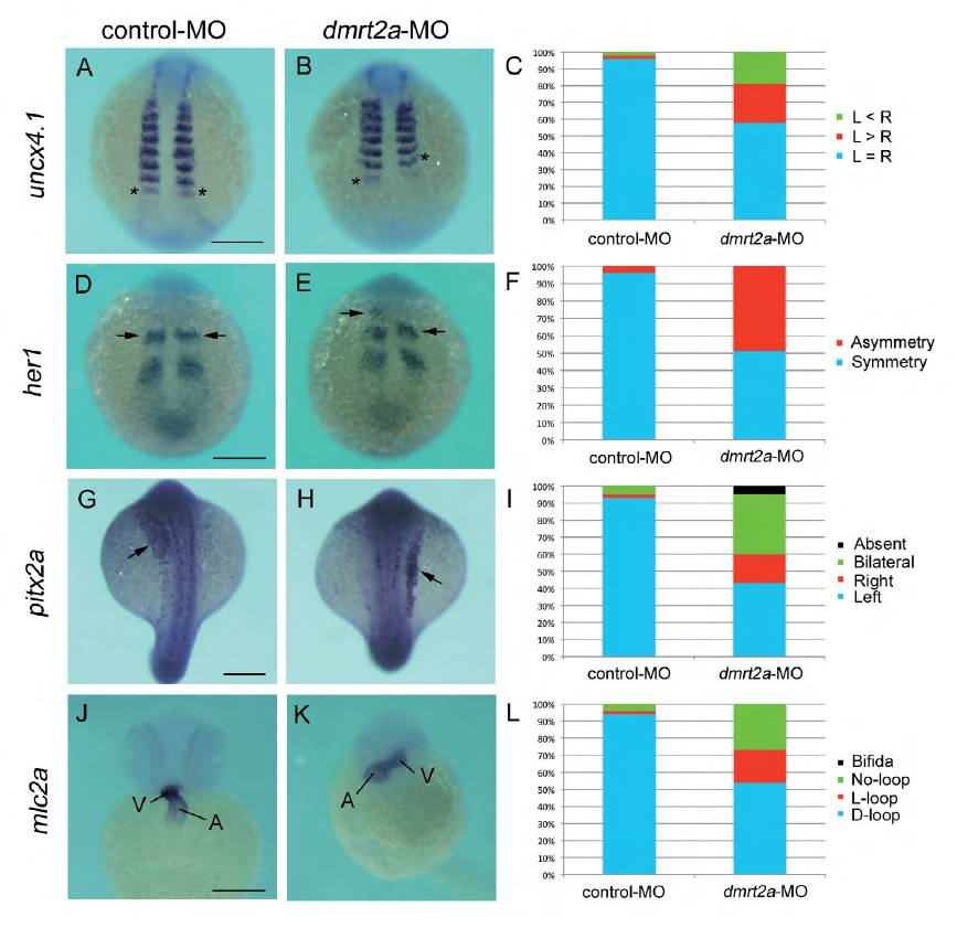
Fig. S2 Knockdown of dmrt2a yields defects similar to those seen in celf1-overexpressing embryos. (A,B,D,E,G,H,J,K) In situ hybridization for uncx4.1 (A,B), her1 (D,E), pitx2a (G,H) or mlc2a (J,K) in control- MO-injected (A,D,G,J) or dmrt2a-MO-injected (B,E,H,K) embryos. Asterisks in A and B mark the last-formed somite. Arrows in D, F, G and H indicate the position of the anterior strip of her1 and pitx2a expression in the lateral plate mesoderm, respectively. A, atrium; V, ventricle. Scale bar: 200 μm. (C) Percentages of symmetric (L=R), left-biased (L>R) or right-biased (L<R) asymmetric somitogenesis in embryos injected with control- MO (n=44) or dmrt2a-MO (n=53). (F) Percentages of symmetric and asymmetric her1 oscillation in embryos injected with control-MO (n=48) or dmrt2a-MO (n=51). (I) Percentages of left-sided, right-sided, bilateral, or no (absent) expression of pitx2a in embryos injected with control-MO (n=59) or dmrt2a-MO (n=46). (L) Percentages of D-loop, L-loop, no-loop or cardia bifida of the heart in embryos injected with control-MO (n=58) or dmrt2a-MO (n=78).
Image
Figure Caption
Figure Data
Acknowledgments
This image is the copyrighted work of the attributed author or publisher, and
ZFIN has permission only to display this image to its users.
Additional permissions should be obtained from the applicable author or publisher of the image.
Full text @ Development

