
|
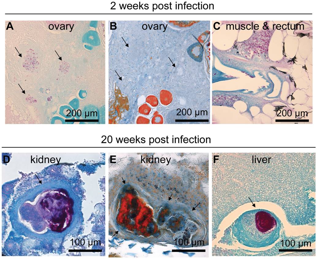
Fig. 2 M. marinum induces the formation of granulomas that mature into well-defined structures during an infection.
In fish infected with a low dose (34±15 cfu) of M. marinum, Ziehl-Neelsen staining at 2 wpi commonly reveals areas with free bacteria (C). Some slightly better formed and restricted areas containing bacteria, here referred to as early granulomas, are also seen (A), but as shown in (B) trichrome staining of the adjacent slide, encapsulation around the mycobacterial lesions is absent at the early stage of infection. At 20 weeks, fish that have survived have mature granulomas (D–F) many of which are multicentric surrounded by a fibrous capsule (D&E). (E) Trichrome staining shows the fibrous capsule in blue (F). The amount of bacteria inside granulomas has increased from the earliest time-points.