
|
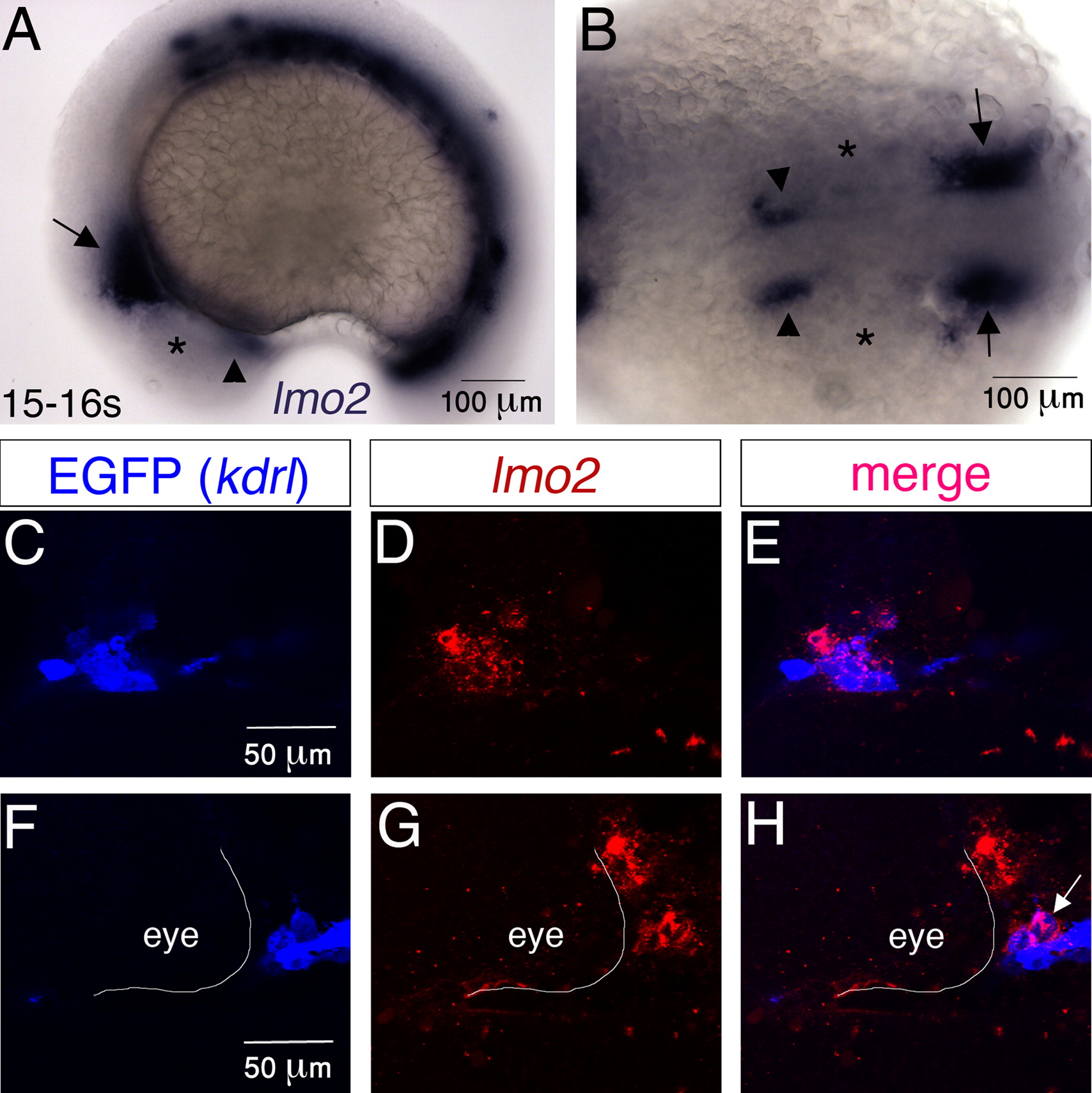
Fig. 3 lmo2 is expressed in the developing head vasculature. (A and B) Whole-mount in situ hybridization showing lmo2 expression in wild-type embryos. Arrowheads and arrows point at the ROC and MOC respectively. Asterisks mark the location of eyes. (C?H) 2 μm single confocal sections of combined in situ hybridization for lmo2 expression (red) and antibody labeling for EGFP (blue) in endothelial precursors in Tg(kdrl:EGFP) embryos. lmo2 is expressed in most ROC (C?E) and in some (arrow) MOC (F?H) endothelial precursors. In (F?H) the posterior and medial borders of the eye are outlined. All images are of 15?16 somite-stage embryos. (A) Lateral view and (B-H) dorsal view, anterior to the left.
Reprinted from Developmental Biology, 369(2), Weiss, O., Kaufman, R., Michaeli, N., and Inbal, A., Abnormal vasculature interferes with optic fissure closure in lmo2 mutant zebrafish embryos, 191-198, Copyright (2012) with permission from Elsevier. Full text @ Dev. Biol.