Image
Figure Caption
Fig. S2
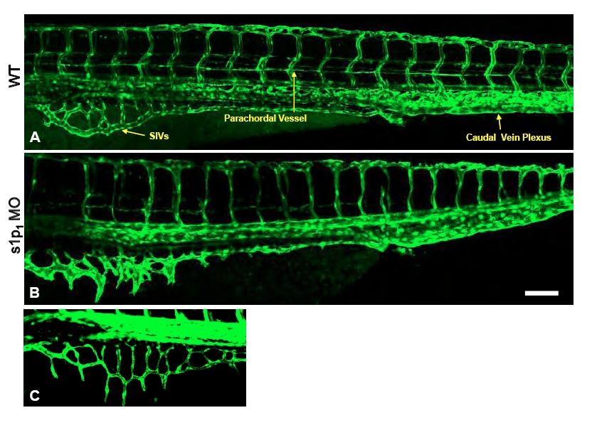
S1p1 knockdown in zebrafish causes defects in blood vessel formation. (A-C) Fluorescence microscopy of control (A) and slp1 MO (B,C) embryos at 3 dpf. Arrows indicate subintestinal vessel (SIV), parachordal vein and caudal vein plexus. SIVs in the slp1 MO embryo exhibit two phenotypes, namely dilated vessels (B) and ectopic sprouting (C). Scale bar: 100 μm.
Figure Data
Acknowledgments
This image is the copyrighted work of the attributed author or publisher, and
ZFIN has permission only to display this image to its users.
Additional permissions should be obtained from the applicable author or publisher of the image.
Full text @ Development

