Fig. S1
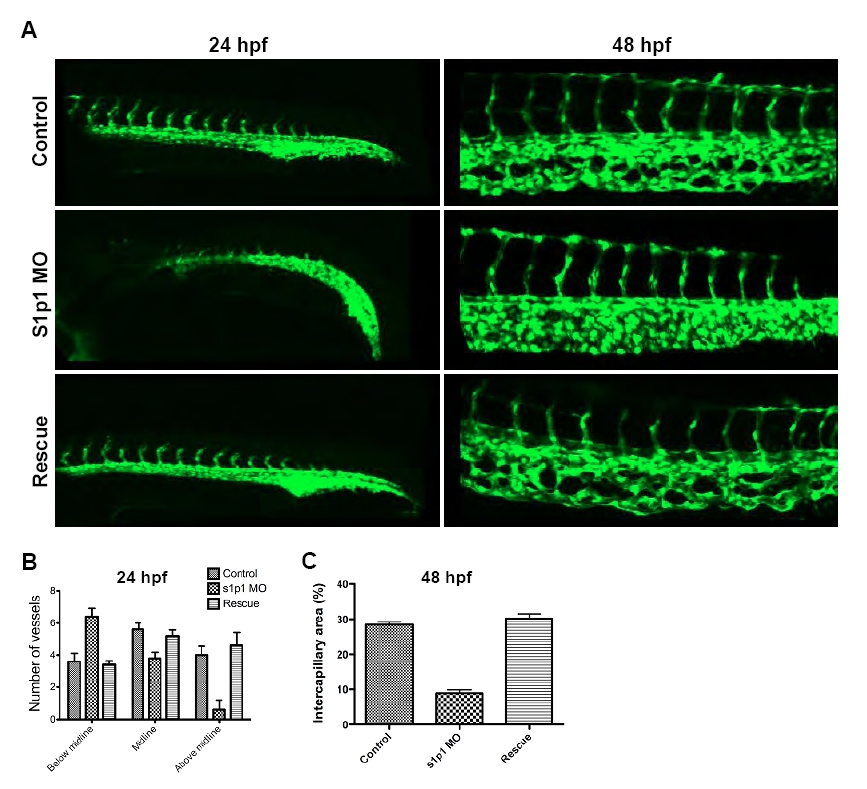
Rescue of the vascular phenotype by s1p1 mRNA injection. (A) Fluorescence microscope images of control (upper panel), s1p1 MO (middle) and s1p1 mRNA-injected morphant (lower panel) zebrafish embryos at 24 (left) and 48 hpf (right). (B) Quantification of ISV location relative to the midline. Both mRNA-injected (rescue experiment) and non-injected s1p1 MO zebrafish were compared with control embryos (n=6 WT, 6 MO and 6 mRNA-injected embryos, 6-8 ISVs from each embryo were analyzed; s1p1 MO: P<0.05; rescue experiment: Pbelow midline -0.74, Pabove midline=0.74; error bars represent s.e.m.) (C) Comparison of the intercapillary area in the CVP between WT, slp1MO and mRNA-injected zebrafish (n=6 in all groups; Ps1p1 MO=0.002, PRescue=0.39, error bars represent s.e.m.).

