
|
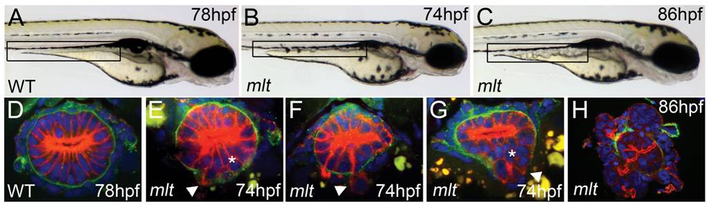
Fig. 1 Intestinal epithelial invasion in mlt larvae.
(A?C) Live images of wild type (WT) and mlt larvae. In WT (A) the posterior intestine forms a smooth cylindrical tube (box), whereas in mlt at 74 hpf the intestinal contour is irregular (B). Cystic expansion of the intestine is evident in 86 hpf mlt larvae (C). (D?H) Histological cross-sections through the posterior intestine of larvae immunostained for laminin (green) and cytokeratin (red). The WT intestine is comprised of a simple epithelial sheet consisting of a single layer of cells, whereas in mlt epithelial stratification (asterisks) and invasive cells that have breached the basement membrane are evident (E?G arrowheads). The initial invasive behavior is followed by expansive growth and loss of epithelial architecture (H).