
|
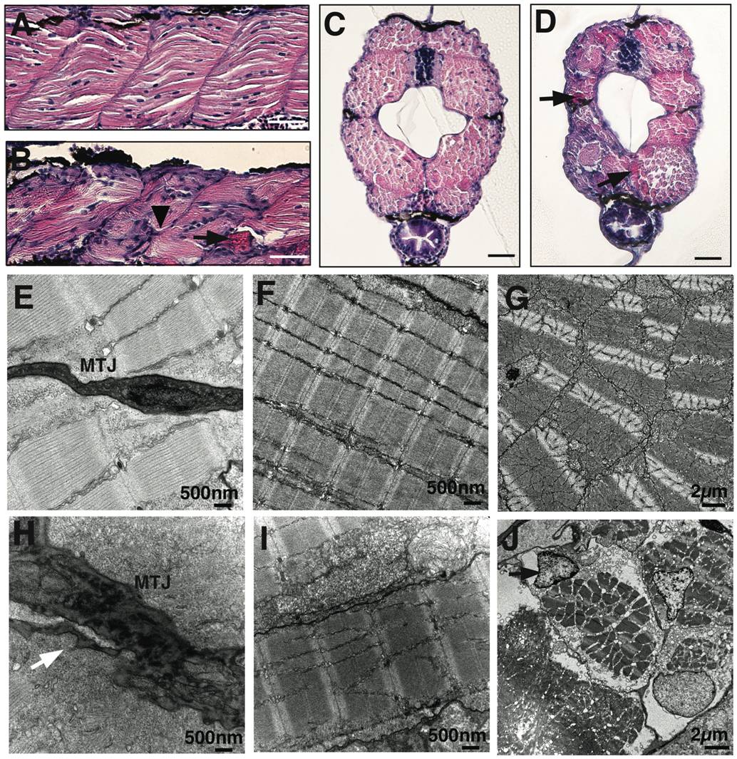
Fig. 2 Abnormal myosepta and myofiber detachment in laminin-α2 deficient lama2cl501/cl501 fish.
(A & B) Hematoxylin and eosin staining of longitudinal sections of wild-type and lama2 mutant fish at 5 dpf. Mutant muscles showed highly disorganized myofibers in the affected somites with irregular myosepta boundaries (arrowhead) and eosin positive detached myofibers (arrow). (C?D) Cross-sections of wild-type and mutation fish also showed smaller myotome and degenerating muscle fibers in lama2 mutant fish at 5 dpf (arrows), bars = 10 μm. (E & H) Electron microscopy showed myofiber detachment from the myotendinous junction (MTJ) in mutant muscles (arrow). (F & I) The myofibers in wild-type muscles attached tightly to the surrounding fibers while mutant muscle displayed large gaps in the extracellular matrix between adjacent fibers and disorganized Z- lines (black arrow) and M-lines (while arrow). (G & J) Defects in extracellular-matrix results in damaged myofibers in the myotome (cross-section). A large number of apoptotic nuclei were observed in the mutant muscles (J, arrow).