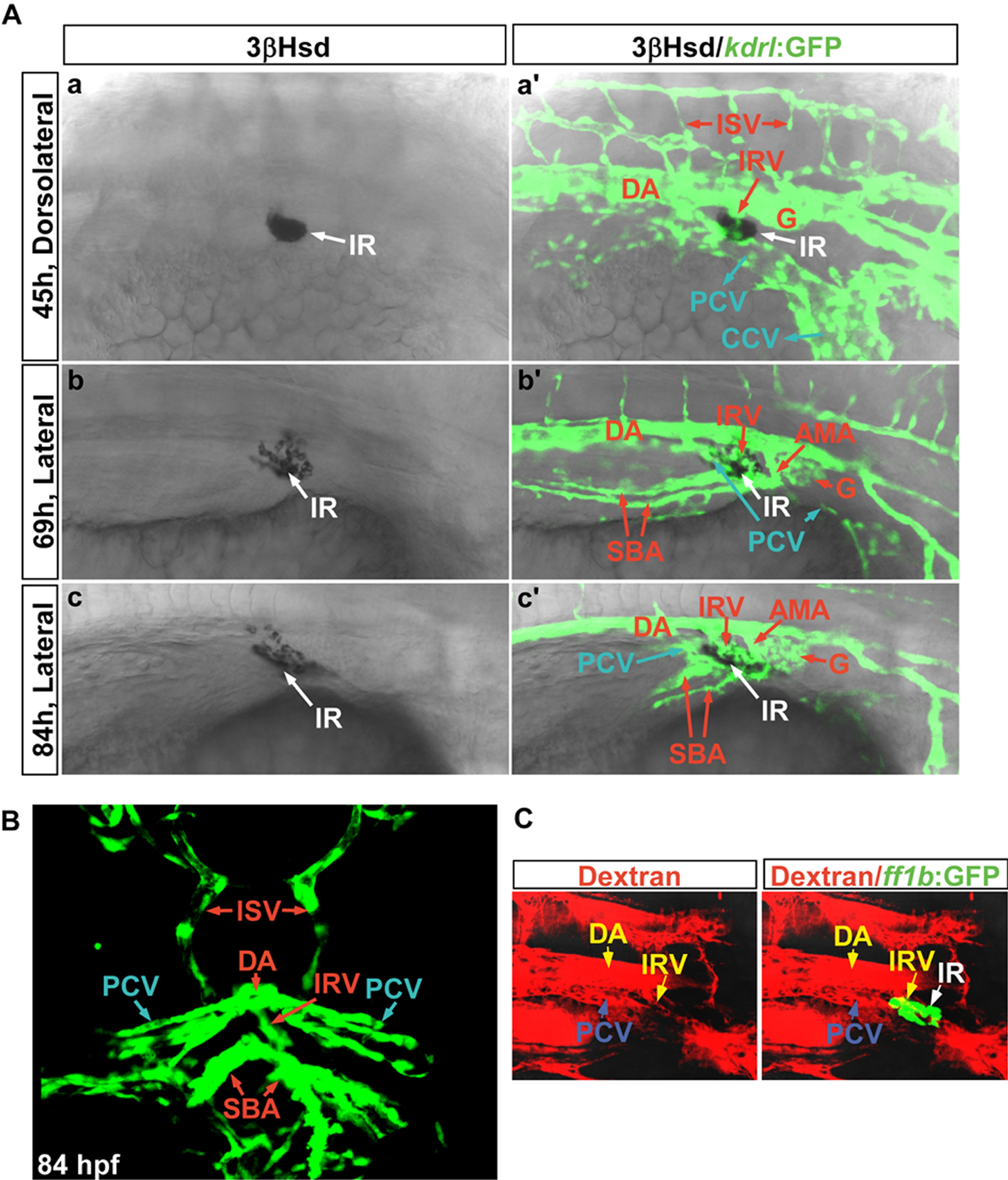
Fig. S1
The IRV is sprouted from the DA and connected to the AMA. (A) Sets of confocal images display the interrenal tissues (IR, white arrows) as detected by 3β-Hsd activity staining, and the neighboring endothelium as labeled by green fluorescence, of Tg(kdrl:EGFP)s843 embryos at 45, 69, and 84 hpf respectively. Panels (a, a′) are dorsolateral while panels (b, b′, c, c′) are lateral views, and all panels are oriented with anterior to the right. The fluorescent image of the vascular pattern for the 45 hpf embryo (a′) was acquired through a projection of a consecutive z-stack encompassing the peri-interrenal area, while single confocal images were shown for the peri-interrenal vascular patterns at 69 and 84 hpf (b′, c′). The IRV was formed caudal to and distinct from the pronephric glomerulus (G) and the AMA. Red and blue arrows denote arterial and venous structures, respectively. (B) The transverse view of the vascular structure neighboring the IRV. The fluorescent image represents a projection of a consecutive z-stack encompassing the IRV, and the more posterior swim bladder artery (SBA) segments. The IRV sprouted from the ventral DA and connected to the AMA segment near which two branches of SBA were branched out. The rotation view of this projection is shown in Video S1. (C) Microangiography by injecting rhodamine-dextran into the blood stream of a Tg(ff1bExon2:GFP) embryo at 3 dpf. The blood circulation through the developing interrenal tissue is established by 3 dpf. Abbreviations: interrenal tissue (IR), dorsal aorta (DA), intersegmental vessel (ISV), interrenal vessel (IRV), glomerulus (G), posterior cardinal vein (PCV), common cardinal vein (CCV), anterior mesenteric artery (AMA), SBA (swim bladder artery).

