
|
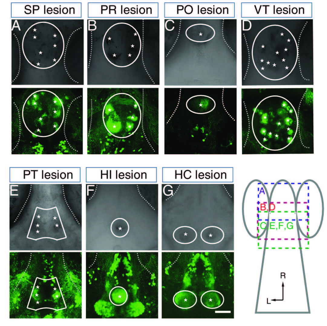
Fig. S6
related to Figure 7. Two-Photon Laser Focal Scanning Effectively Ablates GFP-Positive Cells in ETvmat2:GFP Larvae
(A-G) Brightfield images (top) and GFP-ir signals (bottom) in the subpallium (SP, A), pretectum (PR, B), preoptic area (PO, C), ventral thalamus (VT, D), posterior tubercular (PT, E), intermediate hypothalamus (HI, F) and caudal hypothalamus (HC, G) after two-photon laser-induced focal lesion of the corresponding clusters (white lines). The asterisks mark bulb-like structures, which indicate a successful cell lesion. The dotted lines delineate the position of the eye. Each larva was subjected to laser lesion of only one cluster of neurons. The lesion was performed on ETvmat2:GFP larvae aged at 4 - 5 dpf. The schematic in the bottom-right shows the brain regions, from which images in (A-G) were taken. Dorsal view, rostral is up. L, lateral; R, rostral. Scale: 50 μm.