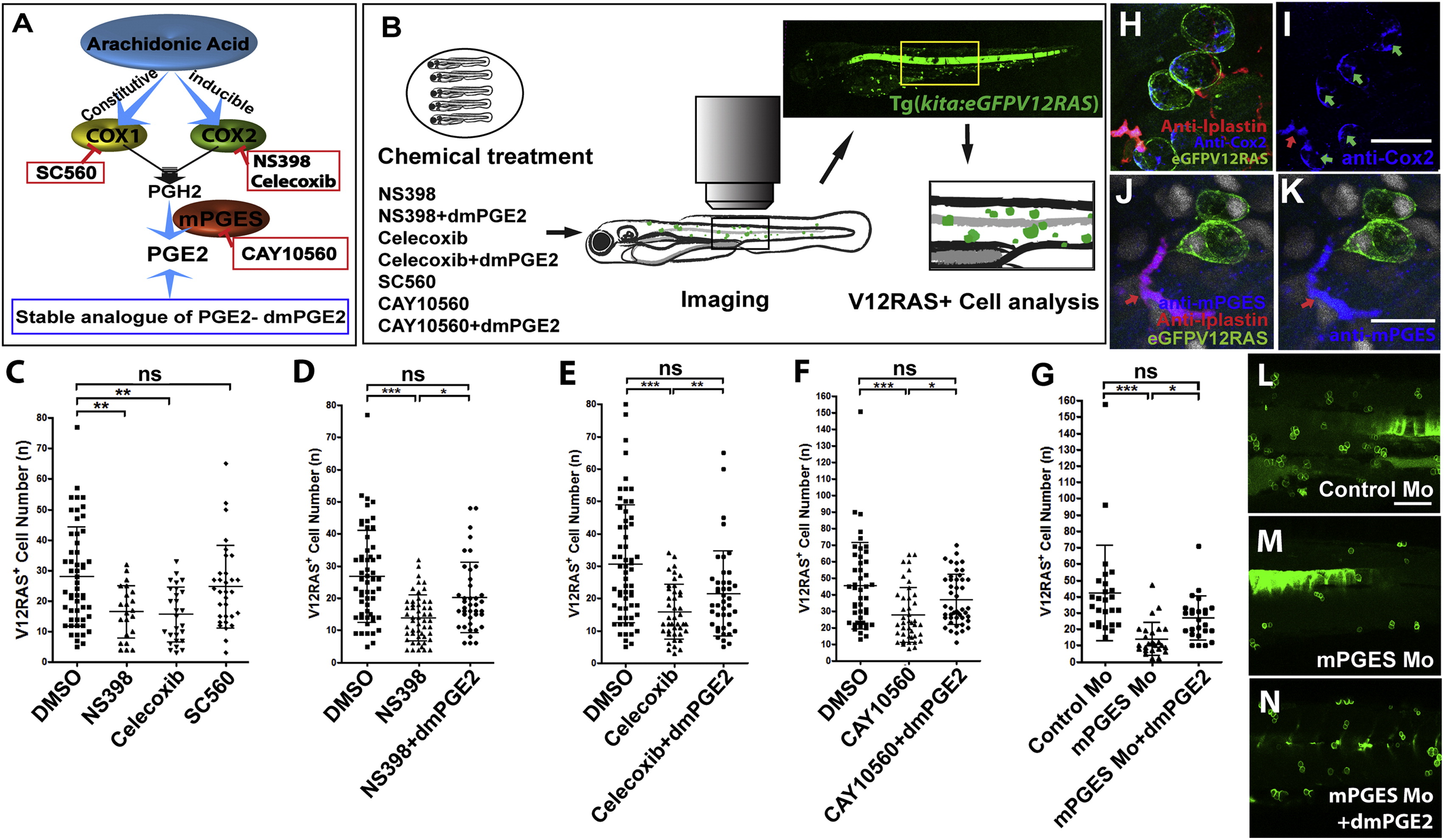
Fig. 1 Blocking COX-2-mPGES-Mediated PGE2 Production Suppresses the Growth of V12RAS+ Transformed Cells In Vivo(A) A schematic representation of PGE2 production through the COX-2 pathway indicates the targets and inhibitors used in this study.(B) A schematic representation of our pharmacological treatment regime and clonal analysis of transformed cells (green) in V12RAS+-cell-bearing larvae. A yellow box indicates the flank skin region in which we quantified alterations in growth of V12RAS+ cells.(C?G) Graphic comparisons of V12RAS+ cell numbers in larval flank skin region after various treatments; (C) larvae treated with DMSO, COX-1 inhibitor, SC-560, and the COX-2 inhibitors NS398 and Celecoxib (p < 0.01, n = 22, 27, and 32, respectively); (D) larvae treated with DMSO, NS398, and NS398 +dmPGE2 (p < 0.001, n = 56, 51, and 40, respectively); (E) larvae treated with DMSO, Celecoxib, and Celecoxib +dmPGE2 (p < 0.001, n = 61, 42, and 42, respectively); (F) larvae treated with DMSO, CAY10560 and CAY10560 +dmPGE2 (p < 0.001, n = 45, 42, and 46, respectively); (G) Control morphants, mPGES morphants, and mPGES morphants rescued with dmPGE2 (p < 0.001, n = 26, 25, and 25, respectively).(H) Immunostaining for COX-2 (blue) indicates that both leukocytes (anti-L-plastin [red]) and V12RASeGFP+ transformed cells (green) express COX-2.(I) Single-channel image of (H), better showing COX-2 expression; green and red arrows indicate transformed cells and leukocytes, respectively.(J) Immunostaining for mPGES (blue) indicates its expression by some leukocytes (anti-L-plastin; red arrow) but not transformed cells (green).(K) Single-channel image of (J).(L?N) Representative images of flank skin regions showing V12RAS+ clones (green) of (L) control morphant, (M) mPGES morphant, and (N) mPGES morphant supplemented with dmPGE2- Scale bars represent 20 μm (H and I), 15 μm (J and K), and 100 μm (L?N). See also Figures S1, S2, and S3.
Image
Figure Caption
Figure Data
Acknowledgments
This image is the copyrighted work of the attributed author or publisher, and
ZFIN has permission only to display this image to its users.
Additional permissions should be obtained from the applicable author or publisher of the image.
Full text @ Curr. Biol.

