
|
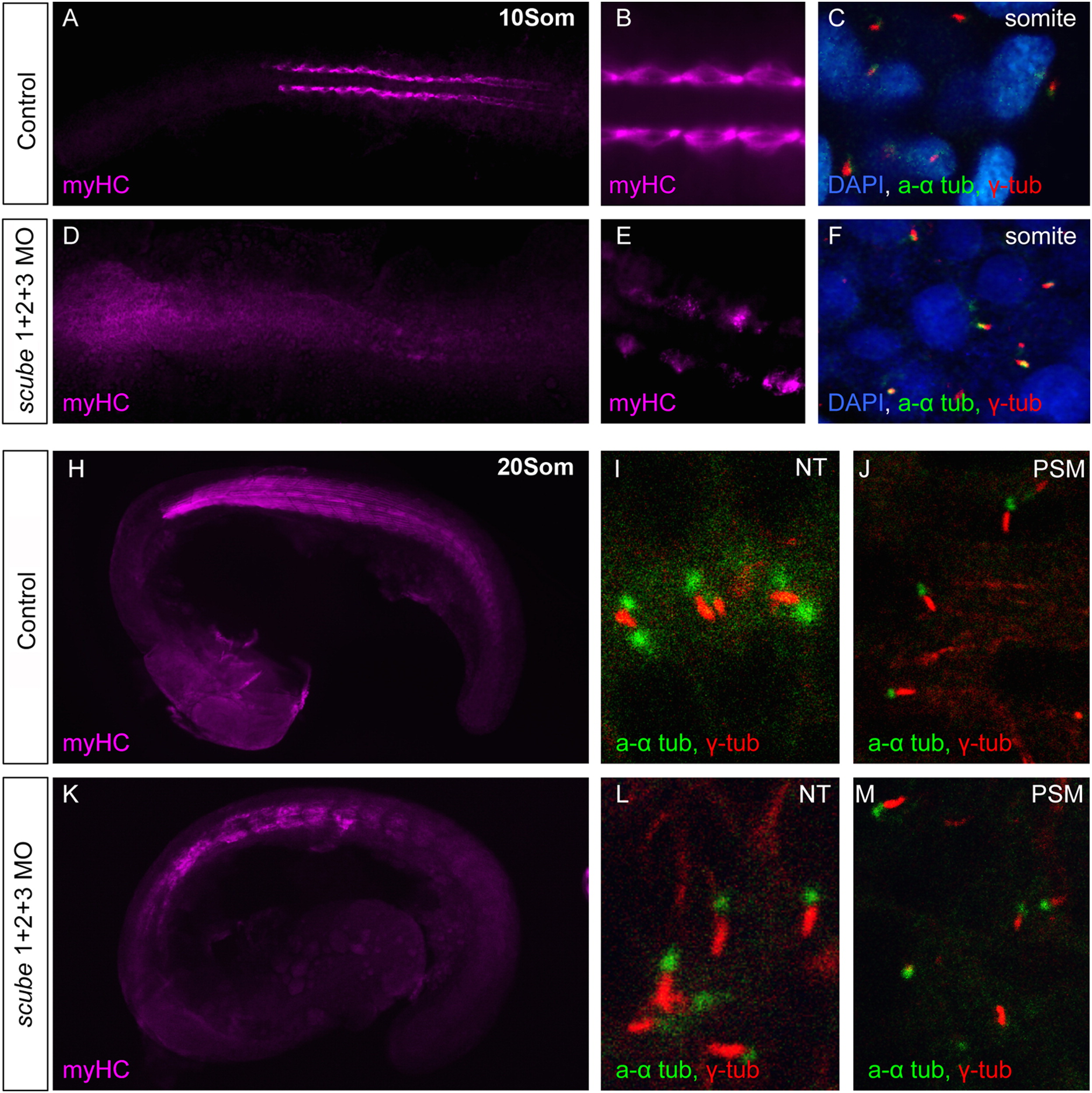
Fig. 6 Scube activity is not required for primary cilia formation. (A–M) Scube triple MO injections do not affect formation of primary cilia. (A–C). Water injected control 10 somite stage embryos display normal slow muscle development visualized by the slow myHC specific antibody F59. (A, B) Primary cilia are present in the somites, marked by acetylated alpha (a α)-tubulin (green) and gamma (γ)-tubulin (red) (Nuclei marked with DAPI) (C). (D–F). 10 somite stage embryos injected with scube triple MO have disrupted slow muscle myogenesis (D, E), yet appear to possess normal primary cilia (F). (H–J). In control 20 somite embryos (H) slow myHC staining shows a well-organised myotome with chevron shaped somites. (I) In wildtype embryos, cilia are visible in the neural tube (NT) and presomitic mesoderm (PSM) (J). (K–M). In scube triple MO embryos at 20 somites, myogenesis is disrupted, with somites displaying a U-shaped morphology (K). Cilia visible in the neural tube (L) and paraxial mesoderm (M) appear normal in scube triple MO embryos.
Reprinted from Developmental Biology, 368(2), Johnson, J.L., Hall, T.E., Dyson, J.M., Sonntag, C., Ayers, K., Berger, S., Gautier, P., Mitchell, C., Hollway, G.E., and Currie, P.D., Scube activity is necessary for Hedgehog signal transduction in vivo, 193-202, Copyright (2012) with permission from Elsevier. Full text @ Dev. Biol.