
|
Fig. S2
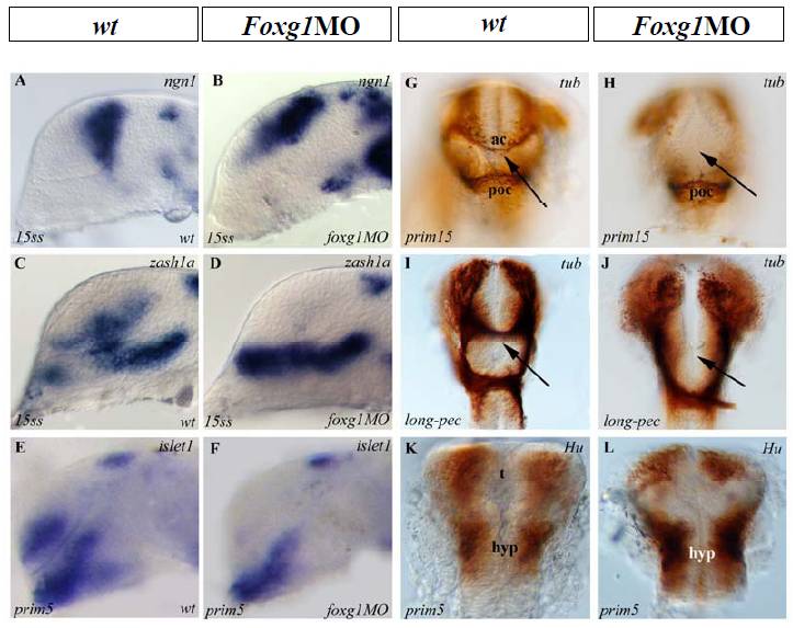
Neuronal Differentiation Is Affected in the Telencephalon of foxg1 Morphant
(A–F) Lateral views, anterior to the left, G-L: frontal views, dorsal top. Developmental stages are indicated at bottom left of each picture. (A) ngn1 expression is detected in the dorsal telencephalic neurons in wt embryos and its expression expands ventrally in foxg1 morphants (B). Ventral neurons detected by expression of zash1a (C) or islet1 (E) are absent in foxg1 morphants (D and F, respectively).
Anti-tubulin immunostaining at later stages show that the anterior commissure (arrows in G and I) is completely absent in foxg1 knockdown embryos (H and J), while the postoptic commissure remains unaffected. Hu immunostaining labelling neurons nuclei in foxg1 morphants (L) in comparison with wt (K). t, telencephalon ; hyp, hypothalamus; d, diencephalon ; ac, anterior commissure ; poc, post-optic commissure.
Reprinted from Developmental Cell, 16(4), Danesin, C., Peres, J.N., Johansson, M., Snowden, V., Cording, A., Papalopulu, N., and Houart, C., Integration of telencephalic Wnt and hedgehog signaling center activities by Foxg1, 576-587, Copyright (2009) with permission from Elsevier. Full text @ Dev. Cell