IMAGE

Fig. S2

ID
ZDB-IMAGE-120725-3
Source
Figures for Sieger et al., 2012
Image
Figure Caption

Fig. S2

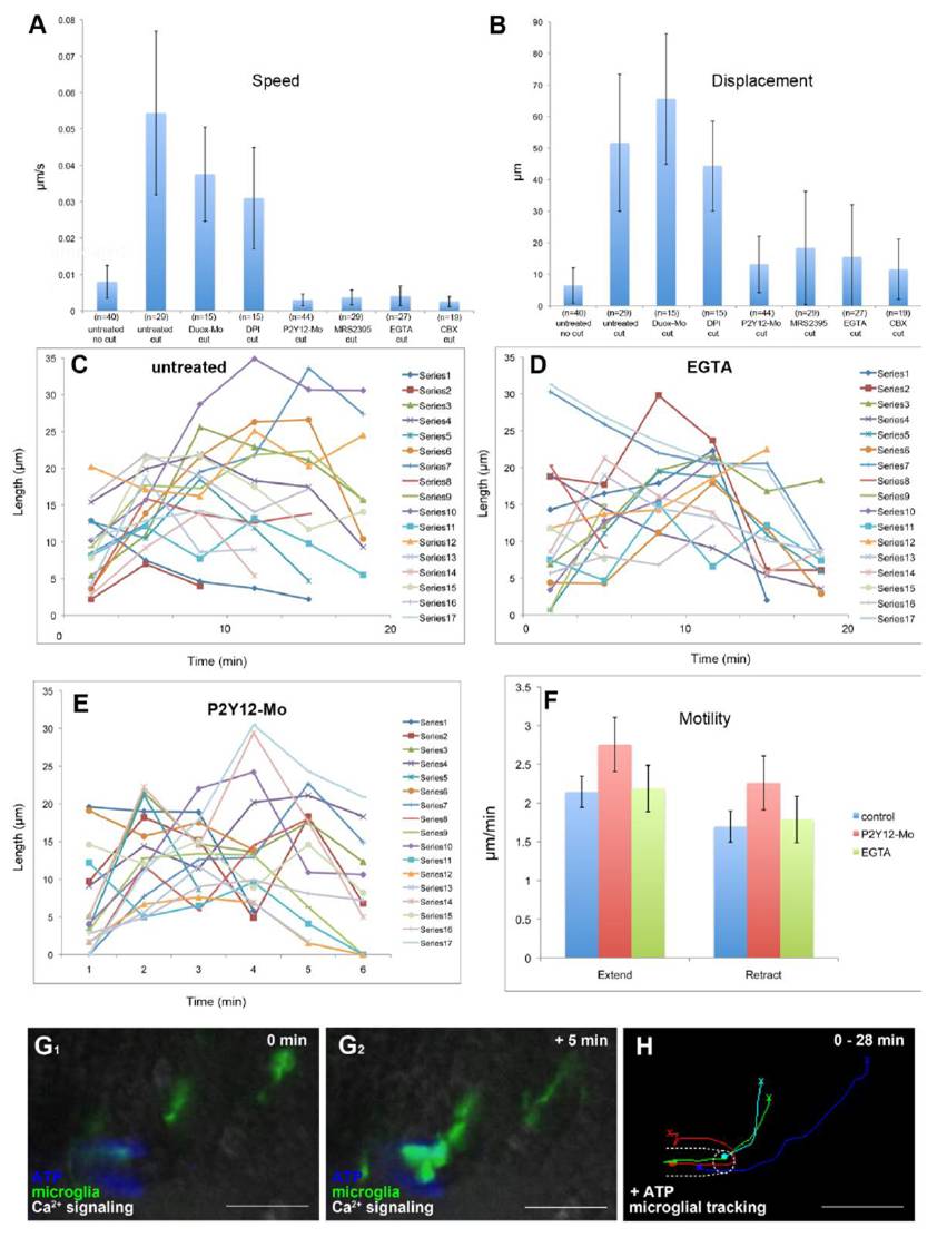
Microglial baseline motility is normal in P2Y12 knockdown and EGTA treated brains, related to Figure 4

(A) Speed of microglial movement (μm/s) upon central injury in untreated embryos compared to speed of movement upon central injury after Duox-Mo injection, DPI treatment, P2Y12-Mo injection, MRS2395, EGTA and CBX treatment. Error bars represent standard deviation. (B) Displacement of microglia (μm) upon central injury in untreated embryos compared to displacement upon central injury after Duox-Mo injection, DPI treatment, P2Y12-Mo injection, MRS2395, EGTA and CBX treatment. Error bars represent standard deviation. (C-E) Length of individual microglial branches over time in untreated (C), EGTA injected (D) and P2Y12 depleted (E) brains (Mov.S4 right). (F) Speed of microglial extensions and retractions in control, EGTA injected and P2Y12 depleted brains. Error bars show standard deviation. (G1 and G2) Microglia (pU1::Gal4-UAS-TagRFP) before (G1) and 5 min after (G2) ATP injection into brain (Mov.S8). (H) Cell tracking of microglia moving towards an ATP point source. The starting point of individual tracks is marked with an X, the end point is marked with a dot. Scale bars always: 20 μm. Images (G1 and G2) were done using an Andor Spinning Disk Confocal with a 20x/NA0.7 objective.

Acknowledgments
This image is the copyrighted work of the attributed author or publisher, and ZFIN has permission only to display this image to its users. Additional permissions should be obtained from the applicable author or publisher of the image.

Reprinted from Developmental Cell, 22(6), Sieger, D., Moritz, C., Ziegenhals, T., Prykhozhij, S., and Peri, F., Long-Range Ca(2+) Waves Transmit Brain-Damage Signals to Microglia, 1138-1148, Copyright (2012) with permission from Elsevier. Full text @ Dev. Cell