
|
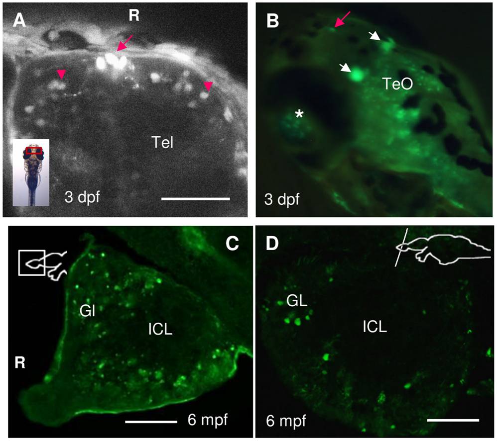
Fig. 2 Expression of spon1b:GFP in the olfactory bulbs (OB) and telencephalon (Tel) of larval and adult zebrafish.
A. Dorsal Tel of 3 dpf embryo, showing individual cells of the OB (red arrowheads), and a cluster of three oval midline cells at the OB-Tel boundary (red arrow). Confocal z-stack image. B. Latero-dorsal view of live larva, at 3 dpf, with robust spon1b:GFP expression in the oval cells of the Tel-OB boundary (red arrow), in the paired habenular nuclei (white arrows), in individual cells of the TeO, and in the eye, with retinal ganglion cells visible through the lens (asterisk). C-D. Sagittal (B) and coronal (C) sections of the adult OB showing spon1b-positive cells in the outer glomerular cell layers (GL), but not in the inner cell layers (ICL). R: rostral end, in A & C. Scale bars: A: 200 μm; C-D: 50 μm.