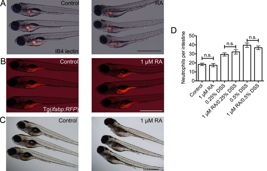
Fig. S5 Doses of exogenous retinoic acid used in this study do not overtly affect intestinal patterning. (A) Representative images of whole mount IB4 lectin staining of the oesophageal-intestinal bulb junction in control and 1 μM RA-treated larvae. (B) Comparison of RFP expression in Tg(ifabp:RFP)as200 control and 1 μM RA-treated larvae. (C) Comparison of neutral red endocytosis by mid-intestinal epithelium in control and 1 μM RA-treated larvae. (D) Enumeration of intestinal neutrophils in larvae co-treated with 1 μM RA and DSS from 3 dpf (n≥30, two biological replicates). Error bars indicate S.E.M., P values determined by ANOVA. All scale bars indicate 1 mm.
Image
Figure Caption
Acknowledgments
This image is the copyrighted work of the attributed author or publisher, and
ZFIN has permission only to display this image to its users.
Additional permissions should be obtained from the applicable author or publisher of the image.
Full text @ Dis. Model. Mech.

