
|
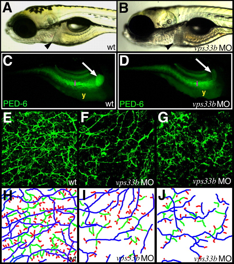
Fig. 3
vps33b knockdown disrupts zebrafish intrahepatic biliary development. (A,B) Left lateral views of 5 dpf wild-type (A) and vps33b (B) morpholino-injected larvae. Liver size (black arrowheads) is comparable in these larvae. (C,D) Right lateral fluorescent images of 6 dpf wild-type (C) and vps33b morpholino injected larvae (D) following ingestion of the PED-6 lipid reporter. Gallbladder fluorescence (white arrow) is decreased in morpholino-injected (D) larva relative to wild-type larva (C). i, intestinal fluorescence; y, endogenous yolk fluorescence. (E-G) Confocal projections through the liver of 5 dpf wild-type (E) and vps33b (F,G) larvae processed for keratin 18 immunohistochemistry. There are fewer bile ducts in F than in E; ducts are sparse, with fewer interconnecting ducts and terminal ductules. (H-J) Colorized schematics of bile ducts from E-G. Long ducts depicted in blue, interconnecting ducts in green and terminal ductules in red.