
|
Fig. S1
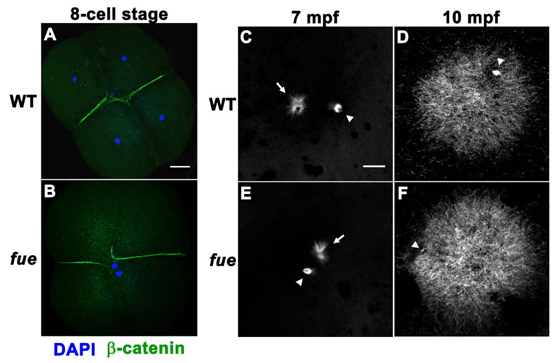
fue Embryos Fail to Undergo Pronuclear Migration and Fusion although the Sperm Aster Is Intact, Related to Figure 1
(A) DAPI (blue) and β-catenin (green) immunolabeling of wild-type embryos at the 4-cell stage.
(B) Same labeling as (A) in fue embryos shows that male and female pronuclei do not fuse though cytokinetic divisions proceed normally resulting in anucleate blastomeres. Scale bar in (A) represents 75 μm and applies to (A,B).
(C-F) Immunolabeling of embryos with anti-α-tubulin antibody to visualize the sperm aster. The sperm aster is of similar size and morphology in both wild-type and fue embryos at 7 and 10 mpf. Arrowheads indicate residual meiotic spindle microtubules while arrows point to the sperm aster. Scale bar in (C) represents 20 μm and applies to (C-F).