Fig. 2
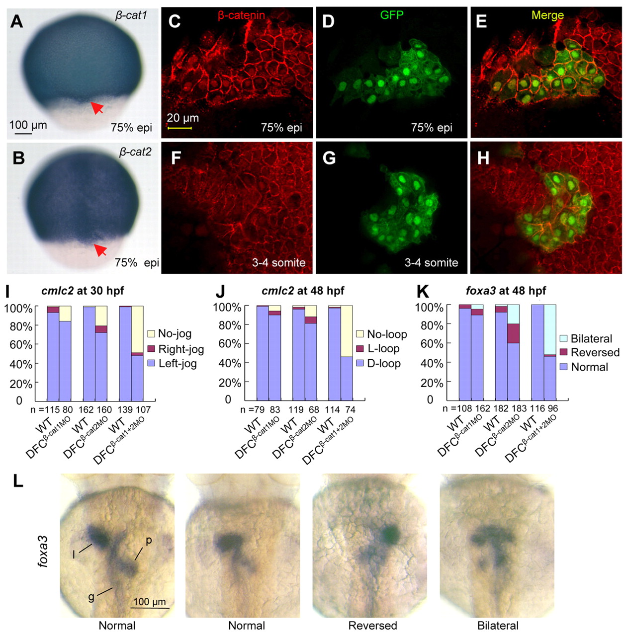
Knockdown of ctnnb1 and ctnnb2 in DFCs causes LR defects. (A,B) ctnnb1 (A) and ctnnb2 (B) expression in DFCs were detected by in situ hybridization. (C-H) Tg(sox17:GFP) transgenic embryos at 75% epiboly (C-E) and the three- to four-somite (F-H) stages were immunostained using anti-β-catenin and anti-GFP antibodies for detection of nuclear β-catenin in DFCs and KV cells. (I-K) Statistical data for the expression of indicated LR markers following midblastula injection. (L) The representative images show foxa3 expression in liver (l), pancreas (g) and gut (g) at 48 hpf. DFCs knockdown of ctnnb1 or ctnnb2 did not block liver and pancreas formation.

