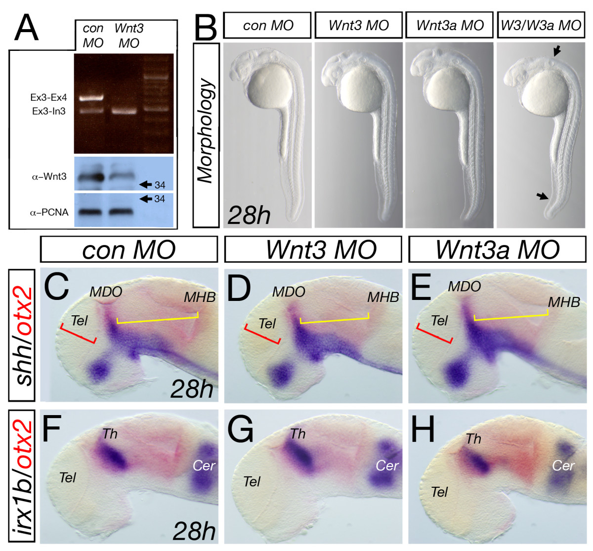
Fig. 3
Analysis of knock-down of Wnt3 and Wnt3a. Injection of wnt3 MO leads to a decrease in splicing of mRNA at the exon3-exon4 boundary and an increase in non-spliced RNA product visualized by amplification at the exon3-intron3 boundary and subsequently to a reduction of Wnt3 protein in morphant embryos at 28 hpf (A). Knock-down of either Wnt3 or Wnt3a leads to an indistinguishable morphology to the control Morpholino-injected embryos whereas the double morphant embryos show a significant alteration in brain morphology, a kinked tail and a smaller otic vesicle (B, arrows). Similarly, analysis of marker genes in the forebrain midbrain area shows no alteration in control, wnt3 or wnt3a morphant embryos (C-H). Cer, cerebellum.

