
|
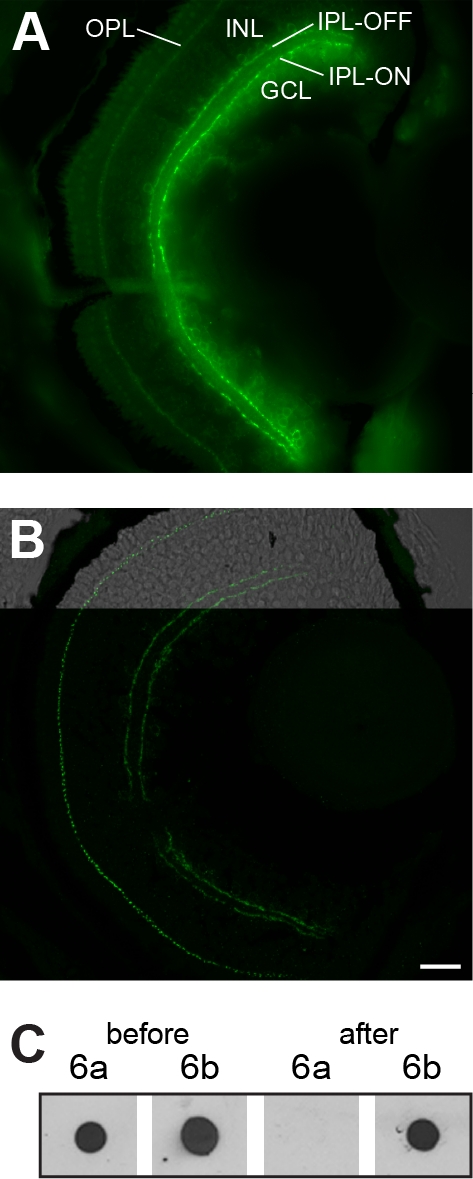
Fig. S2
Cross-absorbance of the mGluR6b antibody. A: Immunohistochemistry image of a cross section through a 5 day old larval retina stained with the original mGluR6b antibody (1:200). B: Confocal image of a larval retina at 5 dpf stained with the cross-absorbed mGluR6b antibody (1:150). For further description see Figure 3. Scale bar in B = 20 μm (applies for A and B). C: Dot-blot analysis showing the increased specificity of the cross-absorbed mGluR6b antibody. 1 �g of mGluR6a (6a) and mGluR6b (6b) epitopes were pipetted on nitrocellulose membranes (0.45 μm; Bio-Rad, Reinach, Switzerland) and incubated with the non cross-absorbed and the cross-absorbed antibodies. Following cross-absorbing the epitope of the mGluR6a is not recognized anymore.