Image
Figure Caption
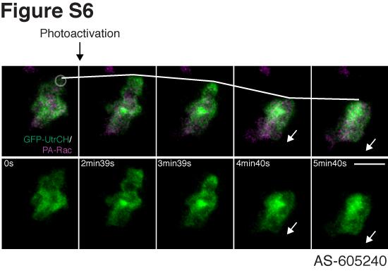
Fig. S6 Transient activation of Rac with PI3K inhibition induces accumulation of stable F-actin at the protrusion, which contracts and pushes the cell body to the opposite direction (related to Figure 6). Rac was photoactivated once at the edge of a cell treated with the PI(3)Kγ-specific inhibitor AS-605240 (movie S13C). After stopping photoactivation, stable F-actin accumulates at the protrusion induced by Rac activation, which contracts and pushes the cell body to the opposite direction. The white arrows indicate direction of cell movement. Scale bar, 10 μm.
Acknowledgments
This image is the copyrighted work of the attributed author or publisher, and
ZFIN has permission only to display this image to its users.
Additional permissions should be obtained from the applicable author or publisher of the image.
Reprinted from Developmental Cell, 18(2), Yoo, S.K., Deng, Q., Cavnar, P.J., Wu, Y.I., Hahn, K.M., and Huttenlocher, A., Differential Regulation of Protrusion and Polarity by PI(3)K during Neutrophil Motility in Live Zebrafish, 226-236, Copyright (2010) with permission from Elsevier. Full text @ Dev. Cell

