
|
Fig. S3
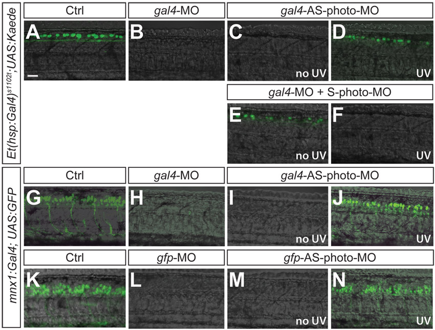
GFP-expression in embryos injected with gal4- or gfp-MOs. (A-F) Lateral view of Et(-1.5hsp70l:Gal4-VP16)s1102t,(UAS-E1b:Kaede)s1999t embryos at 26 hpf injected with gal4-MO. No GFP expression is seen in a Gal4-MO-injected embryo (B) whereas control embryo (A) shows GFP expression in dorsally located Rohon-Beard neurons. (C) gal4-AS-photo-MO without UV blocks GFP expression. (D) Normal GFP expression in embryo exposed to UV at 30% epiboly. (E) gal4-MO/gal4-S-photo-MO leads to normal GFP expression. (F) After UV at 30% epiboly exposure GFP expression is absent. (G-N) Lateral views of Tg(mnx1:gal4, UAS:GFP) embryo at 26 hpf. (G) GFP expression in motoneurons in control embryos. (H,L) GFP expression is blocked in gal4-MO (H) and gfp-MO (L) injected embryos as well as in gal4-AS-photo-MO (I) and gfp-AS-photo-MO (M) injected embryos without UV exposure. (J,N) Restored GFP expression in embryos exposed to UV at 30% epiboly. Scale bar: 30 μm.