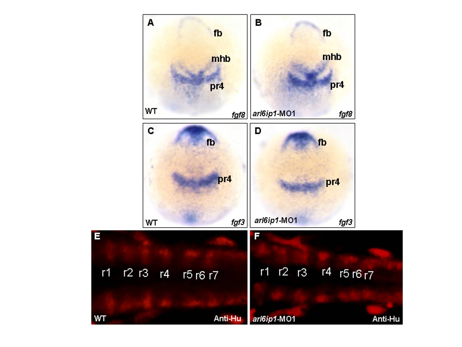
Fig. S2 The arl6ip1-MO1-injected embryos do not appear to have defective patterning in hindbrain. Wild-type (WT; A, C, E) and arl6ip1-knockdown (MO; B, D, F) embryos, either at 3-somite stage (3ss) (A-D) or at 24 hpf (E, F), were observed at dorsal view. (A-D) Neither fgf3 expression nor fgf8 expression in the arl6ip1 morphants was distinguishable from that of wild-type embryos (A vs. B; C vs. D). (E, F) Dorsal views of 24 hpf embryos processed for anti-Hu immunofluorescence staining (IFA) to reveal hindbrain segmentation. Similar to WT embryos, arl6ip1-knockdown embryos showed normal r1-r7 segmentation. fb, forebrain; mhb, midbrain-hindbrain border; pr4, premature rhombomere 4; r1?r7, rhombomere 1?7.
Image
Figure Caption
Acknowledgments
This image is the copyrighted work of the attributed author or publisher, and
ZFIN has permission only to display this image to its users.
Additional permissions should be obtained from the applicable author or publisher of the image.
Full text @ PLoS One

