Fig. S1
Characterization of dysferlin expression and morpholino-injected embryos.
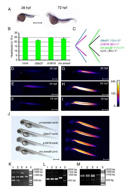
(A) In situ hybridization of dysferlin (dysf) illustrates muscle specific expression at 28 hpf (hours post-fertilization) and at 72 hpf. (B) i36e37 morphants show a slight decrease in heart function, whereas no significant difference was observed for dysf e18i18 or annexin A6 (anxa6) morphants at 72 hpf. (C) Myoseptal angles in dysf i36e37 and anxa6 morphants were significantly different from controls. (D-F) Birefringence imaging over a 48 hour period in a single dysf i36e37 morphant. An initial increase in birefringence could be detected until 54 hpf stage, followed by a drastic loss of muscle structure in the next 24 hours. (G-I) Birefringence imaging in a single uninjected WT embryo. Gradual increase in muscle birefringence could be observed over the 48 hour period. (J) Five-mismatch control morpholinos injected at one cell stage did not induce any phenotypic difference in 3 day old animals. Muscle birefringence (right image) was unaltered. (K-M) Molecular characterization of splicing pattern in morpholino-injected embryos. Total RNA was extracted from single embryos and subjected to RT-PCR (lanes 1-3, morphants; lane 4 control embryo; lane 5, DNA size markers). All PCR products were additionally verified by sequencing. (K) i36e37 morpholino induced removal of exons 37-38 (asterisk), eliminating 80 amino acids, including 32 amino acids of the C2E domain. Alternatively, addition of intron 36 occurred (two asterisks), introducing a premature stop codon after 1393 amino acids. (L) e18i18 morpholino led to 78 bp deletion inside dysf (amino acids 610-635). (M) Anxa6 morpholino caused removal of 94 bp exon, leading to premature stop codon and truncation of the protein after 177 amino acids. Orientation of embryos: anterior left, dorsal up. Scale bar, 500 μm.
Reprinted from Developmental Cell, 22(3), Roostalu, U., and Strähle, U., In Vivo imaging of molecular interactions at damaged sarcolemma, 515-529, Copyright (2012) with permission from Elsevier. Full text @ Dev. Cell

