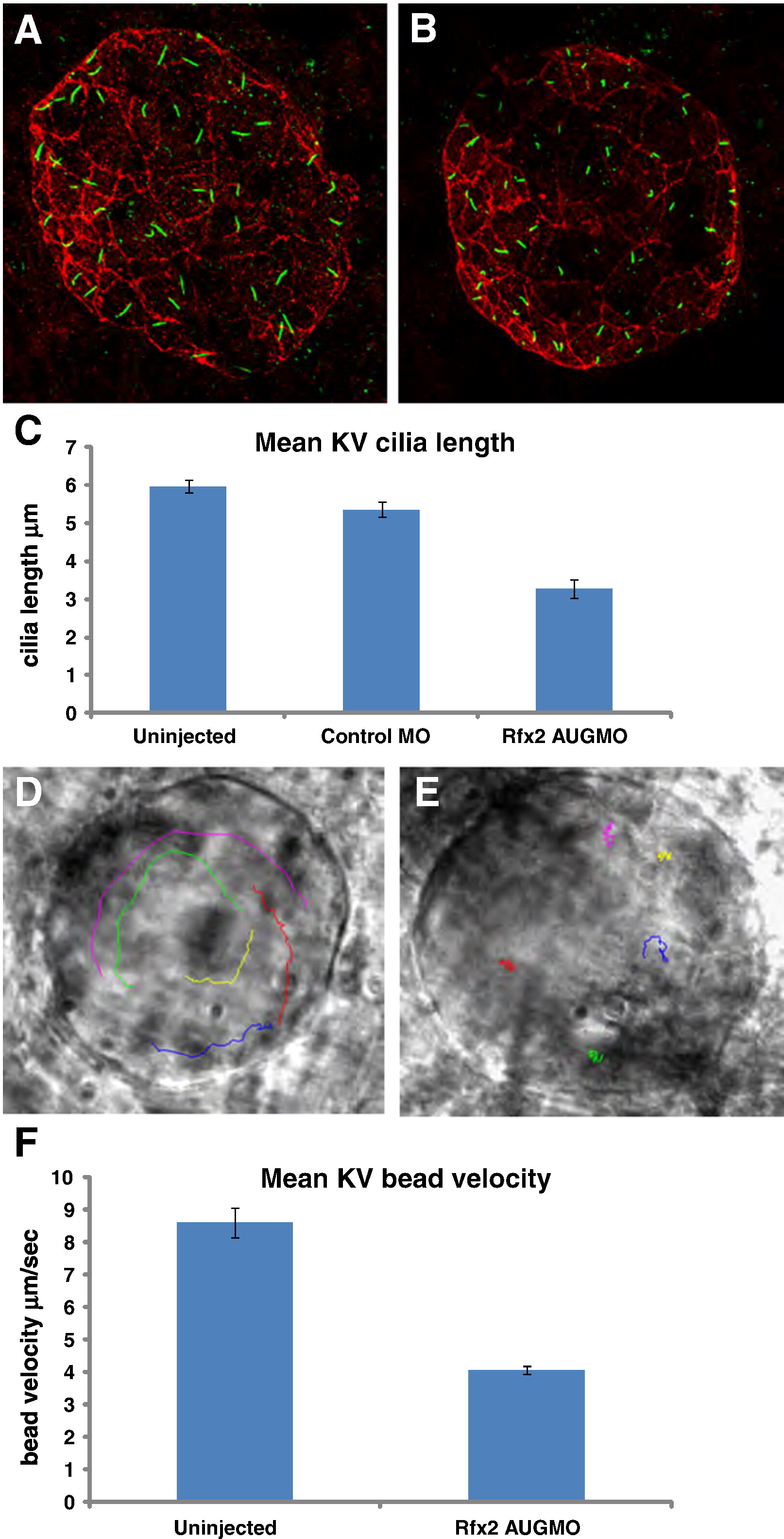
Fig. 4 Rfx2 knockdown disrupts ciliogenesis in KV and KV fluid flow. Cilia within Kupffer′s vesicle of 8?9 somite-stage embryos injected with control morpholino averaged 5.34 μm in length (A) while those in embryos injected with rfx2 AUGMO were significantly shorter, averaging 3.28 μm in length (B,). (C) Histogram of mean cilia length of KV cilia in un-injected, control MO and rfx2 AUGMO embryos (9 embryos each, analysis by one-way ANOVA, error bars are standard error) Fluorescent beads injected into KV of 8 somite-stage control embryos move in a counterclockwise direction (D; Supplemental Video 1A). In rfx2 AUGMO embryos there is no net directional flow and injected beads follow a random trajectory, often reversing direction (E; Supplemental Video 1B). The embryos in panels D, E are oriented with the embryonic notochord in the lower right-hand corner of the image. (F) Histogram of mean bead velocities in un-injected control embryos and rfx2 AUGMO embryos (6 control embryos, 7 rfx2AUGMO; 5 beads tracked in each, analysis by Student′s two tailed t-test, error bars are standard error).
Reprinted from Developmental Biology, 363(1), Bisgrove, B.W., Makova, S., Yost, H.J., and Brueckner, M., RFX2 is essential in the ciliated organ of asymmetry and an RFX2 transgene identifies a population of ciliated cells sufficient for fluid flow, 166-178, Copyright (2012) with permission from Elsevier. Full text @ Dev. Biol.

