
|
Fig. 6
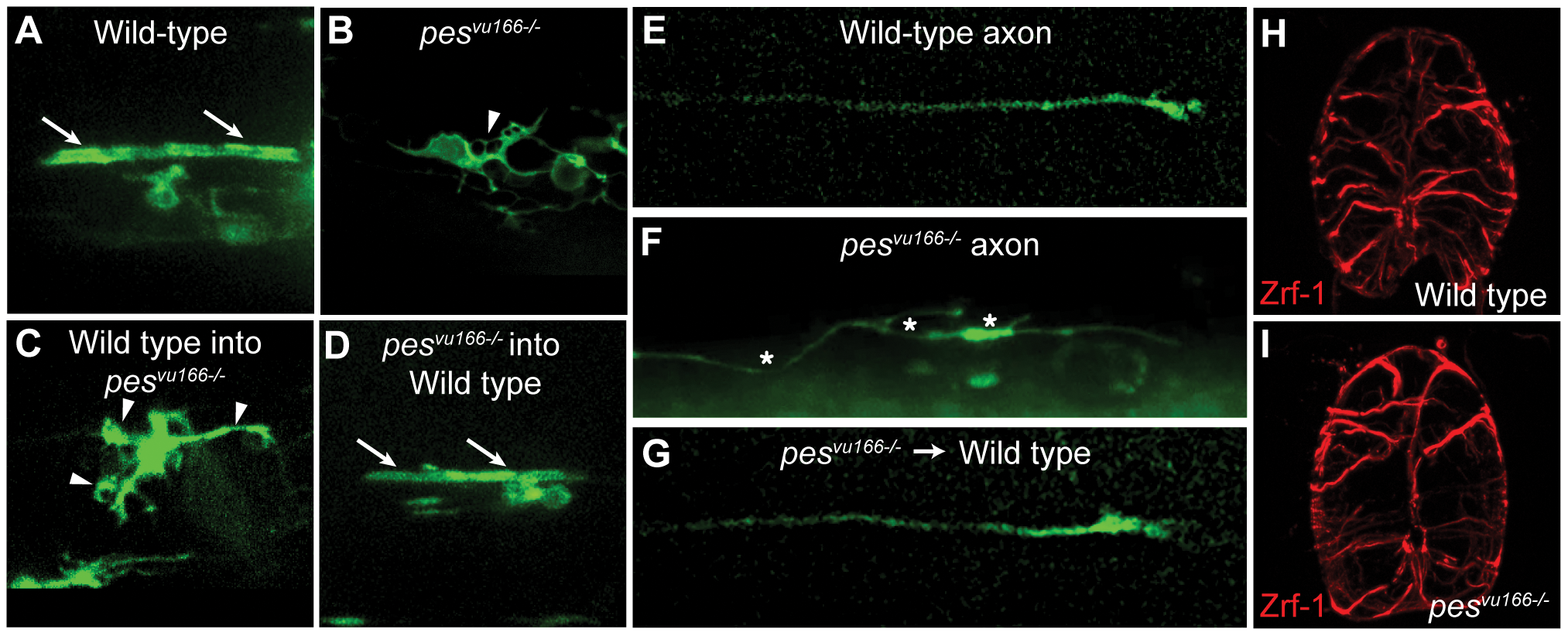
Loss of pesvu166 function results in nonautonomous defects in oligodendrocyte morphology and axogenesis.
(A?G) Lateral views of spinal cords in 3 dpf larvae. Expression from Tg(nkx2.2a:mGFP) reporter marks oligodendrocytes and axons. (A) Oligodendrocyte with normal axon wrapping morphology (arrows) in wild-type larva. (B) Oligodendrocyte with disorganized membrane morphology (arrowhead) and no evidence of axon wrapping in pesvu166 -/- larva. (C) Genetically wild-type oligodendrocyte transplanted into a pesvu166 -/- larva had abnormal membrane extensions and no axon wrapping. (D) Genetically mutant oligodendrocyte transplanted into a wild-type larva wrapped axons normally. (E) Axon in wild-type larva. (F) Axon in pesvu166 -/- larva showing wandering trajectory, abnormal forking and growth cone collapse (asterisks *). (G) Genetically mutant axon transplanted into a wild-type larva had normal trajectory and morphology. (H,I) Transverse sections through the spinal cord of 3 dpf wild-type (H) and pesvu166 -/- (I) larvae showing Zrf-1+ radial glia.