Fig. 5
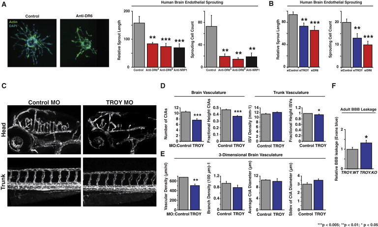
DR6 and TROY Are Required for Proper CNS Vascular Density and Barrier Function through Brain Endothelial Angiogenic Sprouting in a Cell-Autonomous Fashion (A) Anti-DR6 function-blocking antibodies reduce HBMEC sprouting. Actin is green, nuclei are blue (left). Results representative of at least three independent experiments are shown. Quantification of sprout length (middle) and number of sprouting cells (right) are shown. (B) siRNA-mediated knockdown of DR6 and TROY inhibits HBMEC sprouting. (C) Translation-blocking morpholino-mediated TROY knockdown leads to CNS-specific angiogenic defects. Each Tg(kdrl:egfp) zebrafish embryo was treated with 4 ng morpholino. Results representative of at least three independent experiments are shown. (D) 2D quantification of 3 dpf zebrafish vasculature. n = 36 (control_MO), n = 25 (TROY_MO). (E) 3D quantification of 3 dpf zebrafish hindbrain vasculature. n = 5 (control_MO), n = 6 (TROY_MO). (F) Evans blue leakage assay detects barrier defects in adult mice. n = 16 (TROY.WT), n = 18 (TROY.KO). Data are presented as the mean ± SEM (***p < 0.005; **p < 0.01; *p < 0.05). See also Figure S5 and mmc7VIDEO and mmc8VIDEO.
Reprinted from Developmental Cell, 22(2), Tam, S.J., Richmond, D.L., Kaminker, J.S., Modrusan, Z., Martin-McNulty, B., Cao, T.C., Weimer, R.M., Carano, R.A., van Bruggen, N., and Watts, R.J., Death Receptors DR6 and TROY Regulate Brain Vascular Development, 403-417, Copyright (2012) with permission from Elsevier. Full text @ Dev. Cell

